Archive for December, 2011

Rhabdomyolysis – Symptoms, Treatment, Causes, Diagnosis, Pathophysiology

Dec 30 2011 Published by under Diseases & Conditions

What is Rhabdomyolysis?

Rhabdomyolysis is an illness that is a result of speedy break down of the skeletal muscle that releases the myoglobin into blood. This myoglobin is a protein that carries oxygen and can be found generally in the muscle. It is not normally released in the bloodstream because the myoglobin will be filtered by the kidneys for excretion. The kidneys don’t usually filter them because it has big molecular component and it can cause toxicity to the renal tubular epithelium of the kidneys. The myoglobin is not totally toxic in nature but it has a ferrihemate part that releases free hydroxyl radicals which are highly nephrotoxic thus may lead to acute renal failure.

Presence of myoglobin in the blood is abnormal but a relevant mark that detects a muscle injury especially if it involves the heart’s muscle.
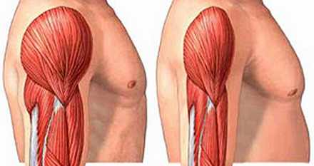
Image of Rhabdomyolysis

Picture of rhabdomyolysis showing muscle mass loss (right)

Pathophysiology

When muscles cells are damage directly by trauma or by cell metabolism, the fluids will shift from extracellular compartment (bloodstream) into the cells which cause it to swell and put pressure against the nerves and the surrounding tissues. This pressure occludes the blood supply to the muscle and may damage the muscle cells.

The shifting of fluid to the cells causes less circulating fluid in the bloodstream, low blood pressure, and possibly, shock. There will be less blood flow to the kidneys to supply the nutrients and the oxygen it needed to function. It creates further harm to the kidneys resulting to renal failure when Calcium binds to the damaged muscles cells. There begins series of persistent muscle contractions as well as depletion of Adenosine Triphosphate that give rise to the fragmentation of the cell and release of the proteins to the bloodstream. The proteins such as the myoglobin may accumulate in the tubules of the kidney and is highly nephrotoxic. The myoglobin attaches itself to the Tamm-Horsfal protein found in the kidneys’ nephrons to form solid aggregates that occludes the blood flow. The condition worsens when there’s presence of high levels of uric acids that further damage the kidney cells.

Causes &Risk factors

The main cause of the Rhabdomyolysis is the muscle injury like:

Direct muscle injury

  • Severe extensive burns
  • Physical torture and abuse
  • Car accidents
  • Prolonged surgery
  • Crush Syndrome
  • Electrocution

Severe muscle contractions due to

  •  Intense physical work-out
  •  Prolonged involuntary muscle contractions during seizures and epileptic episodes
  •  Delirium Tremens in persons who suffer alcohol withdrawal
  •  Tetanus – muscle spasms such as Risus Sardonicus (facial spasms), lockjaw, and Opisthotonos.

Metabolism

During the shortage of the Adenosine Triphosphate in generating energy in muscle, the body compensates by breaking down the fat and protein reserves in the muscle to be used as ATP.

Decrease in the level of electrolytes such as phosphates, potassium, calcium that are essential for the production of ATP.

  • Hyperthermia and heat stroke
  • Hypothermia
  • Prolonged immobilization – patients who have been lying on bed for a long time and the hard surface of the bed may put weight against the muscles that can be enough to bring injury to the muscles.
  • Drug abuse such as cocaine, amphetamines, and heroine
  • Medications such as statins (simvastatin, lovastatin, atorvastatin), anesthesia, psychiatric medications (antidepressants, hypnotics, antilipemic agents)
  • Bacterial and Viral Infections by Epstein-Barr Virus, Herpes, HIV, Influenza A virus
  • Autoimmune diseases such as polymyositis, sickle-cell disease and dermatomyositis

Symptoms & Signs

Mild symptoms

  1. Muscle tenderness, pain, swelling
  2. Muscle weakness
  3. Dark cola-colored urine

Severe symptoms

When there is acute renal failure the severe symptoms starts, which are like

  • Swelling of the upper and lower extremities
  • Compartment Syndrome – the fluids from the bloodstream shifts to surrounding tissues like the lungs (causes shortness of breath), muscles that may lead to low blood pressure and shock.
  • Electrolyte Imbalance
  • Decreased and/or absent urine output
  • Lethargy
  • Disorientation and confusion
  • Irregular heart rate and rhythm
  • Comatose

Diagnosis

In detecting the presence of Rhabdomyolysis, it usually suspected to persons who had trauma or accident and the diagnosis of the Rhabdomyolysis begins by physical examination and the history of past and recent events done by the health care practitioner. It is to assess the person’s history if there are obvious trauma present, the severity of the damage, medication use, or if there is an underlying disease that causes it. This relevant information will help to focus what will be the management for the illness.

In addition to that, there are various diagnostic examinations that are indicated to be performed for persons who are suspected with Rhabdomyolysis:

    • Blood tests 

such as the complete blood count, prothrombin time (PT), partial thromboplastin time (PTT), and electrolytes.

  • An electrolyte such as Potassium tends – to increase in the severe form of Rhabdomyolysis. There is also a decrease in Calcium levels because calcium binds to the damage cells.
  • Electrocardiography (ECG) reflects the elevated Potassium level by the presence of T wave and broad QRS Complex.
  • Kidney function tests such as the Blood Urea Nitrogen (BUN) and Creatinine are done to measure the kidneys’ ability to filter the blood and excrete the body waste via the urine.
  • Urinalysis, in the other hand, is also helpful to confirm if myoglobin is present in the urine which is an abnormal result.
  • Creatinine Kinase (CK) test is the most reliable diagnostic examination in confirming the presence of Rhabdomyolysis. The Creatinine Kinase is an enzyme released by the muscle if there is an injury done in the muscles. It is usually high and continues to rise during the first twelve (12) hours after the muscle injury and then falls gradually after 1 to 3 days. It is a sensitive marker to confirm the Rhabdomyolysis because once the Creatinine Kinase level failed to decrease 3 days after the injury, it is indicated that the Rhabdomyolysis is present. Since it is present in the cardiac muscle and the brain, the laboratory exam can distinguish which muscle cell is affected by performing CK-MM for skeletal muscles and CK-MB for the cardiac muscle.
  • Imaging Studies such as CT Scans and X-Ray are done to visualize the suspected fractures, possible presence of cerebral edema, the extension of brain trauma, and possible seizure activities.

Treatment

Medical Care

Medical Care for the person with Rhabdomyolysis depends on the severity of the illness:

  • If the patient has the mild form of Rhabdomyolysis, home treatment takes place by doing rehydration. Drinking a lot of fluids and rest will work.
  • On the other hand, home therapy is not applicable for those who have the severe form of the Rhabdomyolysis for it requires intensive monitoring of the electrolyte levels, acid-base balance, urine pH levels, ECG tracings, and the metabolic activities.
  • Intravenous infusion is done to avoid the symptoms of shock caused by the Compartment Syndrome. Infusion of large amount of saline increases the flow of fluid to the kidneys, dilute the toxins such as the myoglobin, and correct the levels of electrolytes and the acid-base balance. Mannitol and Furosemide is used to enhance the osmosis and urine production to reduce the kidney damage.
  • It is important to monitor the urine output of the patient hourly by inserting an indwelling foley catheter.
  • Correction of the levels of Potassium and Calcium to remove the risk of developing fatal dysrhythmmia, ventricular tachycardia, and/or ventricular fibrillation. It also removes the risk of alkalinization of urine that possibly causes acute tubular necrosis (ATN).
  • Transfusion of blood products such as the fresh frozen plasma (FFP), platelets, fresh whole blood (FWB) if the complication of Disseminated Intravascular Coagulation is present.
  • To preserve the kidney function is one of the focuses of the treatment and dialysis may be required for the patient with renal failure, consistent hyperkalemia and electrolyte imbalance, metabolic acidosis, congestive heart failure, and pulmonary edema to filter the unnecessary body wastes such as the myoglobin that the failed kidneys can’t filter.
  • The person should avoid medications that are nephrotoxic that will aggravate the renal failure.

Surgical Care

  1. This is necessary depends on the severity of the Compartment Syndrome and pressure to perform the fasciotomy. Normally, high pressure of >30 mm Hg is required to perform the fasciotomy.
  2. Orthopedic and surgical treatment are done to correct the fractures caused by the trauma.

Prognosis

The patient’s chance to recover from Rhabdomyolysis is favorable as long as it is detected on the early stage and given the proper treatment to avoid the further damage to the kidney.  Mortality rate for Rhabdomyolysis is approximately 5% but it still depends on the health status of the patient, the severity of the muscle trauma, the kidney function, and presence of complication. It increases to 20% if kidney failure is present.

Complications

There are three complications the Rhabdomyolysis causes:

1. Compartment Syndrome

It is a condition that happens during an injury that causes shifting of the fluids to third spaces and the muscles which causes swelling and increase in pressure within the surrounding tissues where leads to occlusion of the blood supply to the muscles and compression of the nerves, thus loss of blood supply and possible death of the muscle and the nerve.

2. Disseminated Intravascular Coagulation

A pathological condition where bleeding occurs because of blood coagulation impairment. It is detected by diagnostic examinations such as the PT and PTT.

3. Kidney Failure

The myoglobin released by the muscle to the bloodstream will be filtered by the kidneys and has ferrihemate part that is highly nephrotoxic that may cause renal failure. In addition to that, the shifting of fluid from the bloodstream to the intracellular compartment may cause decreased blood flow to the kidneys. It deprives it of blood, oxygen, and nutrients that are essential for kidney function.

Prevention

Rhabdomyolysis is prevented to be developed if the proper management is implemented after the injury. It involves the

  1. adequate hydration
  2. exercising the range of motion to aid the muscle trauma and a lot time for resting

No responses yet

Altitude Sickness

Dec 25 2011 Published by under Diseases & Conditions

What is Altitude Sickness?

Altitude sickness is known for its different names such as Acosta disease, Acute Mountain Sickness (AMS), Soroche, hypobaropathy or puna. Altitude sickness is a disorder that occurs when the person who is traveling on high elevation such as the mountains with great speed within 2500 meters (8, 0000 feet). There is an oxygen level decrease as he/she ascends with great speed that it didn’t let the body to adjust to the oxygen level. Even the healthiest person with no history of having any disorder is not exempted in acquiring Altitude Sickness episodes.

Symptoms

Mild symptoms that occur in Acute Mountain Sickness, or Altitude Sickness within the first 2,000 meters which causes common symptoms such as:

  1. Headache
  2. Nausea and Vomiting
  3. Body malaise
  4. Nosebleed or Epistaxis
  5. Dizziness and drowsiness
  6. Insomnia
  7. Appetite loss
  8. Dizziness
  9. Swelling of the upper and lower extremities (peripheral edema)
  10. Rapid pulse
  11. Diarrhea or stomach upset

These symptoms above serve as warning signs that the person has the risk to develop severe symptoms of high altitude cerebral and pulmonary edema:

  1. Severe headache
  2. Confusion and Disorientation
  3. Hallucination
  4. Unstable gait and body coordination
  5. Fainting and Unconsciousness
  6. Fever
  7. Shortness of Breath
  8. Persistent cough with pinkish frothy sputum
  9. Paleness of the skin
  10. Comatose

Causes

What causes the person to have altitude sickness? When the person ascended in a rapid pace and stayed on a place where there is low oxygen level, the person will manifest different signs and symptoms affecting the cognition and coordination that can be fatal and deadly to the person. How do you know that the place has less oxygen compared to the normal sea level? It is known that the higher the place is, the less the air pressure in the area. It is a fact that the oxygen level in sea level areas is around 21% and as the person goes up, about 5,500 meters, the oxygen level in the air is reduced to half. The human body isn’t that capable to adjust quickly in low levels of oxygen because primarily, the oxygen is one of the vital components of air to keep the body functioning, alive and healthy. In Altitude Sickness, the increase in altitudes has effect on person’s fluids: it leaks out of the blood vessels which accumulate to spaces where it is not allowed such as the lungs and the brain. Once it is not attended properly and did not descend the mountains, the person is risking himself to have a serious complication of the Altitude Sickness, or worst, death.

Diagnosis

Altitude Sickness is confirmed and diagnose if the person who climbed an altitude of more than 2, 500 feet and manifested one of the mild symptoms of the Altitude Sickness, it is accurate that the person is having the illness.

If the person still experiences the symptoms, the person must undergo diagnostic exams such as Complete Blood Count, Chest X-Ray, and Urinalysis to rule out possible illnesses of hypothermia or flu (because it has the same symptoms as the Altitude Sickness). In addition to that, Chest X-Ray can confirm the presence of High Altitude Pulmonary Edema (HAPE) and CT-Scan or Magnetic Resonance Imagery for High Altitude Cerebral Edema (HACE).

Treatment

Descending

The best way to treat the Altitude sickness is remove the cause: the increase in altitude with low levels of oxygen. The person must descend in order to meet the levels of oxygen in a specific lower altitude, or if not possible, the person must stay on the place and rest.

Rest and doses of pure Oxygen

It is important for the person to rest to let the body adjust to the level of the altitude and the oxygen in the area and avoid doing any physical activities that will increase the demand for oxygen. If the symptoms persist, the person must descend 300 more meters and intake of prescribed medicines to alleviate the symptoms. In addition to that, it is also important to have an adequate supply of oxygen for the body so having an oxygen tank during such situation will help.

Gamow bag

This can be used, if the person has this in possession, when the descent is impossible. This Gamow bag is an implatable bag with chambers that is large enough to fit one person inside. It is primarily used to aid the patients with severe symptoms.

More importantly, the person must NOT IGNORE THE ALTITUDE SICKNESS SYMPTOMS.

Remedies

Acetazolamide

This is a medication that can be taken during the ascending to the mountains and that will help the person to adjust the ascending altitude and descending levels of oxygen. Its main mechanism is that it prevents and lessens the occurrence of the Altitude Sickness Symptoms and it corrects the chemical imbalance caused by the Altitude sickness in the blood.

Dexamethasone

It is a steroid hormone which is its main mechanism is to suppress the immune system and inflammation. This is a medication that remedies the cerebral edema caused by Altitude Sickness. It reduces the inflammation and swelling of the brain. It is reported that the medication takes effect and reduces the cerebral edema after six hours (approximately).

Pain killers

These medications such as Paracetamol and Ibuprofen are taken for symptoms relief like headache and fever.

Prevention

A person must be prepared before ascending a mountain and take note of the following points that will prevent to acquire Altitude Sickness:

altitude illness pictures

  1. Start and ascend GRADUALLY AND SLOWLY – to avoid shocking the body of low levels of oxygen and have the body adjust to the decreasing level of oxygen at high places (Altitude Acclimatization).
  2. Take rest and limit physical activities – to decrease the body’s demand for oxygen
  3. Limit climbing of 3, 000 feet per day.
  4. Hydration – One must take adequate fluids to keep the body hydrated. Avoid alcohol because it causes dehydration. Take at least four to six liters of water every day.
  5. Sleep low – Never sleep on places higher (no more than 300 meters) than you slept the night before.

No responses yet

Fibromuscular Dysplasia

Dec 21 2011 Published by under Diseases & Conditions

What is Fibromuscular Dysplasia?

Fibromuscular dysplasia can involve the leg arteries or rarely the arteries in other areas of the body. Usually it involves many arteries in the body.

The walls of the arteries are made up of three layers:

  1. Tunica intima or the innermost layer
  2. Tunica media or the middle layer
  3. Adventitia or the outermost layer

Fibromuscular dysplasia is classified based on the involved arterial layer and the lesions’ structure. Based on the classification of fibromuscular dysplasia, the narrowing of the artery is brought about by excessive fibers or muscle parts of the wall of the arteries.

Despite the fact that the classification of fibromuscular dysplasia can only be diagnosed 100% by examining the arterial wall under a microscope following a biopsy or surgery this is unusually done.

Generally it is likely to diagnose the classification of fibromuscular dysplasia depending on the form of the arteries involved in the dye angiogram examination.

Types

Here are the five classification of fibromuscular dysplasia, arranged from the most typical to the least:

1. Medial fibroplasia

75 to 80% of the cases of fibromuscular dysplasia are this classification

involves the middle layer (tunica media)

described by parts of fibrous lesions with aneurysms (bulgy parts)

looks like beads on the string in the dye angiogram (hallmark sign)

2. Intimal fibroplasia

occurs in lower than 10% of all the cases of fibromuscular dysplasia

brought about by fibrous tissue deposits surrounding the innermost layer (tunica intima) of the artery

without beads, smooth, concentric and narrowed appearance in the dye angiogram (hallmark sign)

3. Perimedial fibroplasia

lower than 10% of fibromuscular dysplasia are this classification

huge fibrous tissue deposits found in the adventitia or the outermost layer

irregular and thickened walls of the artery

beads that have a small diameter rather than normal artery (hallmark sign)

intensifies the probability of full obstruction of involved arteries

4. Medial hyperplasia

1 to 2% of lesions are under this classification

brought about by over formation of the muscular cells

no fibrous tissue deposits

the appearance is the same with intimal fibroplasia in the angiogram test

5. Periarterial hyperplasia

unusual less than 1% of the cases are in this classification

brought about by widening of the adventitia

the fibrous deposits spread around the fatty layers

defined by swelling of the artery

Symptoms & Signs

The majority of the people suffering from fibromuscular dysplasia are asymptomatic; however, symptoms can happen if the narrowing is serious enough to obstruct blood circulation on the involved artery.

Symptoms of moderate case of fibromuscular dysplasia in the carotid artery:

  1. headaches
  2. tinnitus (ringing in the ears)
  3. lightheadedness

Symptoms of advanced case of fibromuscular dysplasia in the carotid artery:

  1. stroke
  2. TIA or transient ischemic attack

Symptoms of carotid dissection (tear in the artery)

  1. headache
  2. acute neck pain
  3. stroke in advanced cases
  4. transient ischemic attack in advanced cases

Symptoms of fibromuscular dysplasia in the arteries in the kidney

  1. elevated blood pressure
  2. renal insufficiency
  3. commonly does not lead to renal failure

Causes

The exact cause of fibromuscular dysplasia is not yet known. This condition is most probably caused by many underlying reasons. Some of the reasons that may have a role involve:

Hormonal imbalances

Fibromuscular dysplasia happen mostly in women

Heredity

10% of all the cases are inherited. It may also be brought about by genetic defects affecting the arteries.

Trauma or stress

Trauma or stress especially on the walls of the arteries

Lack of oxygen to the wall of the blood vessel

This happen when the capillaries in the walls of the arteries that provide oxygen get obstructed by fibrous lesions.

Diagnosis

Diagnosis of Fibromuscular dysplasia is done with blood vessel studies. The below are common investigations to diagnose fibromuscular dysplasia

  1. Duplex ultrasound
  2. Angiography
  3. CT Angiography
  4. Magnetic Resonance Angiography
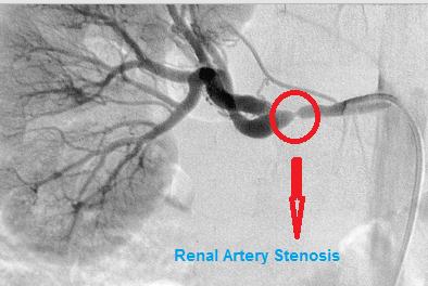
fibromuscular dysplasia angiography
Angiography showing Renal Artery Stenosis in Fibromuscular Dysplasia

Treatment

The following are the treatments for fibromuscular dysplasia:

Medications

When fibromuscular dysplasia does not have any symptoms, it is typically benign and does not need management. For these people, the doctor may give an anti-platelet drug to avoid blood clots. The anti-platelet drug may be given or your doctor may advise that you use aspirin regularly.

People suffering from high blood pressure brought about by fibromuscular dysplasia may be prescribed with anti-hypertensive medications, especially ACE inhibitors.

Know and Control Risk Factors

Risk factors like hypertension, diabetes and elevated cholesterol should be assessed and controlled. People suffering from these problems must undergo CT scan, MRI or duplex ultrasound regularly to monitor the advancement of the disease. This is especially necessary when an aneurysm is confirmed.

Angioplasty

In special cases, percutaneous angioplasty of the arteries of the liver is advised. Same with the method applied to control obstruction in the cardiac arteries, renal angioplasty includes placing of a balloon catheter in the artery at the area of the narrowing or obstruction. The catheter is directed inside the blood vessel with the help of an X-ray. The balloon will be inflated to open the artery and then the balloon catheter is removed.

Putting a stent or a small tube at the area of the obstruction has not been tested to enhance the effectiveness of angioplasty and does not intensify the outcomes. Usually the stent is only used when angioplasty alone is not enough to enhance the blood circulation.

Angioplasty is also advised to those people suffering from fibromuscular dysplasia of the internal carotid artery who suffer from stoke or transient ischemic attack. Stenting may be needed in unusual cases, when people suffering from fibromuscular dysplasia have had vertebral or carotid artery dissection or aneurysm.

Surgery

Reconstructive surgery may be advised for people suffering from complicated fibromuscular dysplasia of the arteries of the kidney or suffering from an aneurysm of the internal carotid or vertebral arteries. Surgery will be based upon the area or the extent of the condition, even though usually it includes removal or bypassed of the involved part of the artery to recover the normal blood circulation.

Prognosis

At present there is no permanent cure for fibromuscular dysplasia. Medicines and angioplasty can lessen the probability of stroke. In unusual cases, aneurysms associated with fibromuscular dysplasia can rupture causing bleeding inside the brain that can lead to stroke, irreversible brain damage or death.

No responses yet

Coagulopathy – Definition, Causes, Symptoms, Treatment, Pictures

Dec 16 2011 Published by under Diseases & Conditions

Coagulopathy Definition

Coagulopathy is an illness where there is an abnormality in the clotting process of the blood. The normal blood clotting process is important to prevent too much blood loss from the body after an injury. When a person accidentally cuts himself, the blood clots within two minutes. The blood solidifies and the clot is made. The blood clot serves as plug on the site of injury that prevents the gushing of the blood out of the body. This blood clotting process is made possible by the existence of adequate number of proteins in the blood such as the albumin, clotting factors, and the platelets. In coagulopathy, this normal process may be happening because of the low or high levels, or worst, absence of the blood protein, clotting factors, and platelets which results to uncontrolled massive bleeding.

 Coagulopathy Causes

Out of many blood disorders, there are causes that make the blood’s normal clotting process impaired:

Low levels of blood proteins and clotting factors

The blood proteins like the Albumin are the one that pulls the blood into and keep it from seeping out of the blood vessels while the clotting or coagulation factors and the platelets are the one that makes the blood clot. Once the clot is formed, it serves as a plug or filling on the opening (the area traumatized) so the blood won’t gush out of the body. Once these normal processes dysfunction, the person might suffer excessive blood loss that may lead to shock and possible death if not properly attended.

Intake of anti-coagulation medication

There are medications that impair the normal clotting process like the Heparin and the Warfarin.

Existing liver problems

All of the clotting factors except Factor VIII, megakaryocytes, and vonWillebrand’s Factor are made in the liver. If the liver is impaired and diseased, there are instances that the liver may produce few clotting factors, or worst, none.

Vitamin K deficiency

Vitamin K, or menaquinone, is a fat-soluble Vitamin produced by the liver that plays a big role on blood coagulation. It is an essential vitamin where proteins are dependent. Without the vitamin K, the coagulation proteins will not be active to form a clot. The Vitamin K is also found in food like green leafy vegetables, cauliflowers, and liver.

Underlying Genetic disorders

There are blood illnesses that such as Haemophilia, von WilleBrand’s disease, Factor II deficiency where the genetic make-up of the person’s blood is lacking of the important clotting or coagulation factors. These disorders are hereditary.

Coagulopathy Symptoms

1. Hypocoagulability

Uncontrolled bleeding externally

  1. Bleeding through the mouth and gums
  2. Bleeding through the anus, vagina, or penis
  3. Frequent epistaxis or nose bleeding
  4. Big rashes and hematomas
  5. Easily bruised when bumped into something hard
  6. Excessive and prolonged menstruation
  7. Pallor

Uncontrolled bleeding internally

  1. Joint bleeding – Painful, swelling, and reduced range of motion of the joint
  2. Presence of blood in the urine or hematuria
  3. Presence of blood in the stool (the color may be black or red, and tarry)
  4. Bloated feeling
  5. Increase in abdominal girth – possible bleeding in the gastrointestinal system

Cerebral Effects

  1. Disorientation
  2. Fainting, or Loss of consciousness
  3. Seizures and Convulsions
  4. Changes in vision or loss of it
  5. Memory loss
  6. Tingling sensation and weakness of the lower and upper extremities
  7. Unbalanced gait and difficulty in walking
  8. Speaking problems

Coagulopathy Pictures

Coagulopathy photos

Coagulopathy images, pics

Coagulopathy features on face

Coagulopathy Types

There are two types of Coagulopathy

1. Hypocoagulability

This is a condition of coagulopathy where there is an unusual uncontrolled bleeding.

2. Hypercoagulability or Thrombophilia

This is a condition where there is an increase in blood coagulation that results to thrombosis. These blood clots are possible to occlude the blood vessels and cause deep vein thrombosis, arterial thrombosis, and pulmonary embolism.

Coagulopathy Diagnosis

Coagulopathy are carefully diagnosed by qualified physician by undergoing test of coagulopathy

  1. Bleeding Time – this is done to know the time (in seconds) the small blood vessels to close and stop the bleeding.
  2. Complete Blood Count with Platelet– this is to know the quantity of Red Blood Cells, White Blood Cells, and Platelet counts.
  3. Fibrinogen Level – this is done to measure the number of protein and clotting factor (Factor 1) that plays a role in blood clotting.
  4. Hageman Factor Assay, Factor 5, Factor 7, Factor 8, and Factor 10 level – This is to know the measurement of the levels of the essential clotting factors important for blood clotting process.
  5. Partial Thromboplastin Time (PTT) – is the blood test that measures the total clotting time per cycle.
  6. Liver Function Test – these are liver examinations such as SGPT and SGOT to know if the liver is functioning and capable of producing the important substances and enzymes for body’s use.

Coagulopathy Treatment

Coagulopathy are treated carefully to avoid massive loss of blood that may be fatal to the person.

  1. Medications such as Aminocaproic Acid that prevents the blood clots to break down, Birth Control contraceptives that help to control the excessive and prolonged menstruation, and Desmopressin Acetate to activate the stored blood-clotting proteins.
  2. Blood and Plasma Infusion – to replace the blood loss.
  3. Blood clotting Factor infusion – this depends to what genetic condition the person has and it is important for them to have this because this is what they are lacking. It is to be replenished to avoid uncontrolled bleeding.

Prevention

There is no total prevention for the coagulopathy to occur if the patient has underlying genetic disorder that leads to coagulopathy. To prevent the uncontrolled massive bleeding, the patient must avoid having himself traumatized even the lightest bump because his blood vessels are sensitive enough to bleed and there is no enough clotting factor to stop the bleeding. Also, if the trauma is inevitable, the patient should have enough supply of his medications as well as the infusion of clotting factors to avoid blood loss.

No responses yet

© 2017 eHealthWall.com. All Rights Reserved. Privacy Policy
This website is for informational purposes only and Is not a substitute for medical advice, diagnosis or treatment.