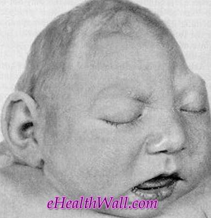
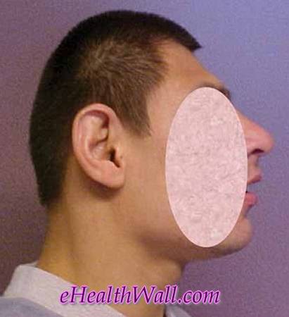
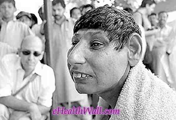
Archive for February, 2012

Gallbladder Pain – Location, Causes and Treatment

Feb 29 2012 Published by under Diseases & Conditions

What is Gallbladder Pain?

Pain is a displeasing sensation that can vary from a discomfort to a point of distress. It may be caused by external or internal stimuli which disrupt the normal functioning of a certain body part or the whole body.

Gallbladder pain is elicited either by the formation or blockage of gallstones in the biliary tracts called cholelithiasis or by an inflammation; a condition called cholecystitis.

Biliary colic is a specific term especially used if the pain is situated at the right upper abdomen, where the gallbladder is located. Although colicky pain literally means it comes and go. In biliary colic it does not come and go, it is constant but may fluctuate from time to time. This should be noted so as to prevent confusion.

Gallbladder location
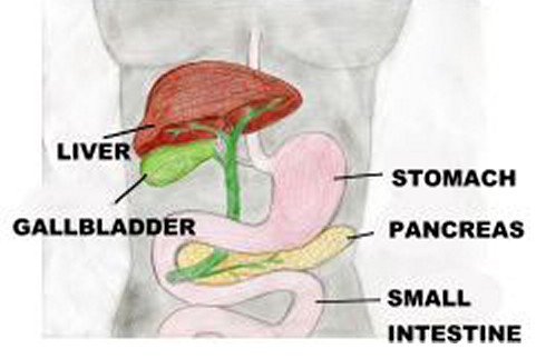
gall bladder location

The gallbladder is a small, pear-shaped saclike organ situated in a depression under the right lobe of the liver. The gallbladder consists of an outer serous peritoneal coat, a middle muscular coat, and an inner mucous membrane that is continuous with the linings of the ducts.

Its primary role is to store bile produced by the liver until the next food intake.

Causes and Risk factors

Cholelithiasis is the existence of calculi in the gallbladder. Usually, this is form by solid constituents. Two major types of gallstones are pigment stones and cholesterol stones. In pigment stones, they are formed by unconjugated pigments in the bile precipitate to form stones while in cholesterol stones, although cholesterol is a normal constituent of the bile, its insolubility to water can cause stone precipitation.

Cholecystitis has 2 subcategories that contribute to its presence. Calculous type is with the presence of gallstones. They block the normal flow of the bile thus causing an inflammation which leads to sever pain. Acalculous type is the absence of gallstones but the pain experienced is the same with the calculous type. This type may be caused by severe trauma, burns or after a major surgical procedure.

The aforementioned conditions play vital roles in the production of pain or biliary colic.

There are certain numbers of factors that can cause gallbladder pain. And there are some people who are more prone to this condition, such as:

  1. Overweight
  2. Thyroid illness
  3. Allergies especially in food
  4. Usage of birth control pills or intake of hormone supplements
  5. Age above 40
  6. Women who already gave birth
  7. High fat, low fiber diet
  8. Diabetes
  9. Crash diet

Signs and Symptoms

Although the patient will experience an excruciating pain in the right upper abdomen that radiates to the back and right shoulder, there are other symptoms that accompany pain like:

  1. Nausea with or without vomiting
  2. Excessive sweating
  3. Generalized body weakness
  4. Light-headedness
  5. Shortness of breath
  6. Abdominal bloating/ belching
  7. Inconsistent bowel movement

Diagnosis

Abdominal X-ray

When a gallbladder blockage is suspected, an abdominal x-ray may be obtained to exclude other causes of the symptom. However, this diagnostic procedure is not just specific to a disease it is very convenient and can immediately give out an idea if the gallbladder is blocked.

If the result is inconclusive, CT scan or MRI should be used; far advanced methods than the traditional one which utilize highly developed equipments.

CT scan stands for computerized tomography scan and this is a method of X-ray slice that takes ‘slice’ of the body cross section or longitudinal section.

MRI, on the other hand, is Magnetic Resonance Imaging does not have X-ray but is very useful in examining the bile ducts.

There are various ways to detect gallbladder disease which is not included in this article. In gallbladder pain, the determination of the cause is the priority and rule out inappropriate diagnoses.

Treatment

When the cause of gallbladder pain has been detected appropriate treatment should be rendered. If the pain is caused by gallstones, there is the surgical and non surgical removal.

Non Surgical

  1. Dissolving gallstones; infusion of a solvent (mono-octanoin or methyl tertiary butyl ether) into the gallbladder. This method is not widely used.
  2. Lithotripsy, this is a non-invasive procedure which uses repeated shock waves directed at the gallstones in the gallbladder or common bile duct to fragment the stones.

Use of medication is also popular in the removal of stones. Ursodeoxycholic acid (UCDA) and chenodeoxycholic acid (chenodoil or CDCA) have been used to dissolve small gallstones composed primarily of cholesterol. Treatment with UCDA can reduce the size of existing stones, dissolve small stones, and prevent new stones from forming.

Nutritional and supportive therapy

Almost all patients who had gallbladder pain caused by stones achieve remission after rest, IV fluids, analgesia, and antibiotic agents.

Diet required is usually limited to low fat liquids. These can include powdered supplements high in protein and carbohydrates stirred into milk. Foods that are well tolerated by the patient are encouraged like rice, lean meats, non gas-forming vegetables and fruits, coffee and tea. The patient should avoid eggs, cream, fatty foods, cheese, rich dressings, gas forming vegetables, and alcohol.

Surgical

Cholecystectomy

The gallbladder is removed through an abdominal incision, usually right subcostal after the cystic duct and the artery are ligated. The procedure is performed after an acute or chronic pain episode.

Laparoscopic Cholecystectomy

This procedure replaced the traditional cholecystectomy that requires an incision to remove the gallbladder. It has become the new standard for therapy for symptomatic gallstones. Advantages of laparoscopic procedure are that the patient does not experience the paralytic ileus that occurs with traditional open surgery and has less post operative abdominal pain. The patient is usually discharge same day or within 2 days and can resume full activity after the surgery.

Conversion to the traditional open abdominal surgery may be necessary if problems are encountered with this advanced procedure.

Although the patient is discharged immediately after surgery, he/she may experience drowsiness, so the patient needs assistance in the first 24-48 hours.
No responses yet

Pompe Disease – Symptoms, Diagnosis and Treatment

Feb 22 2012 Published by under Diseases & Conditions

What is Pompe Disease?

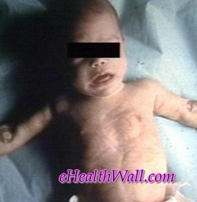
Pompe disease is a neuromuscular kind of disorder that is multisystemic and progressively fatal in nature. It was coined by a Dutch Pathologist named Joannes C. Pompe. It is known as a genetic condition occurring when there is minimal glycogen, which is a type of sugar, stores in the body’s cells.

Pathophysiology

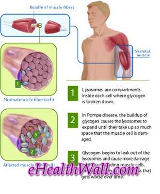
Without glycogen, the function of important muscles such as the heart will be very much affected. With this disease, the GAA or Alpha Glucosidase is oblivious or is decreased significantly.

Hence, excessive glycogens are not broken down and are left to accumulate in the tissues, which shouldn’t be the case for it will result to damage. It is also known as glycogen storage disease type II, AMD, alpha-1,4- Glucosidase deficiency, GSD II or acid maltase deficiency. The effect of this, result to problems that deal with important body organs of the body, such as the heart, the lungs and the muscle per se. This disease will make the muscles weak. The most affected individuals are the infants or children. However, there are occurrences that adults are not exempted from having the disease. The infants or children are the ones who are often reported to have a diagnosis of this disease condition.

Pompe disease pathophysiology picturesImage source – wikipedia.org
Forms

There are actually two forms of this disease: The Juvenile form and the adult form.

1. Juvenile form

With regards to the Juvenile form, it occurs in the primary decade of respiratory difficulties and weakness of the proximal area. It often resembles the dystrophy of the limb-girdle.

2. Adult form

On the other hand, the adult form varies. It may occur within the second to the sixth decade. It is in here where the lower limbs appear to be more severely affected. The respiratory muscles are weak disproportionately. Due to this, some cases may be fatal because it can lead to respiratory failure.

Symptoms & Signs

As mentioned earlier, the pompe disease can affect infants, children and adults alike.

The infants who have this disease will manifest the following symptoms:

  1. weakness of the muscles
  2. enlargement of the liver and heart
  3. breathing problems
  4. unable to eat well
  5. dysphagia or swallowing difficulties
  6. enlarge tongue
  7. has a hard time in holding the head up
  8. no or little weight gain

Meanwhile, the adults are reported to manifest the following symptoms:

  1. having difficulty in climbing or walking in the stairs
  2. generalized weakness in the muscles

Due to the fact that there is accumulation of glycogen in the tissues, the muscles of that organ having increase glycogen will, in time, experience fatigue leading to generalized body weakness. When this happens, the person will have difficulty in engaging in any kind of physical activities. They become drowsy all day long.

They also will encounter difficulty in breathing and in sitting or climbing the stairs, for instance. With regards to infants, their development will be affected. The child or infant shows lack of appetite, experiencing little or no weight gain at all, and may have problems with their muscle development. As a result of having problems in their muscular development, the child may not be able to effectively hold their head up by their own. Aside from that, they will experience muscle symptoms.

The disease itself will progress gradually. They might notice that as the disease progresses, they become more and more weak. They will have difficulty walking, climbing stairs or even standing from a sitting position. Since, they have difficulty walking; they are prone to injuries due to falling. There is also what is called as spine symptoms. What occurs here is during the phase where the muscle weakens, the spine may not be able to fully support the body’s weight. This will result to spinal curvature and will be painful whenever they walk or stand. Lastly, they will have breathing symptoms. They experience episodes of dizziness, labored breathing and fainting.

Diagnosis

To diagnose such disease condition, the symptoms has to be present. However, this first step enough won’t make you directly conclude that it is Pompe. The confirmatory diagnosis for this disease will rely mostly on examination of the Acid Alpha Glucosidase activity which is seen in cultured skin cells of a particular person claiming to manifest its symptoms. Blood test can also be done in adults. In diagnosing, you should keep in mind that the correct confirmation that a person has this kind of disease, the enzyme should be reduced or absent.

Treatment

ERT or Enzyme Replacement Treatment

The present treatment for this disease is ERT or enzyme replacement treatment. Since the disease either lacks, can’t manufacture nor has insufficient supply of the enzyme called Acid Alpha Glucosidase.

Myozyme

While those who are infant or children who also have the disease, are prescribed and treated with Myozyme, which is a lysosomal glycogen specific enzyme.

Lumizyme

Lumizyme, which is an algluosidase alfa medication, which is an FDA approved drug that treats late onset or non-infantile persons diagnosed with this disease condition.

Multidisciplinary Team Approach

In order to effectively treat this disease condition, a team approach is needed. The team members includes speech therapist, genetic counselors, respiratory therapist, occupational therapist, nutritionist or dietician, psychosocial therapist, and finally physical therapist.

Prognosis

When the disease is diagnosed at an early stage in an infant child, disease progresses rapidly, with delay of treatment or without any interventions being done, the prognosis is poor. Chances are, the infant will die. Children and Adults, alike, displayed the rates of the progression of this disease in a more gradual and variable manner.

The prognosis with this kind of disease will depend upon the age of onset. Usually without any treatment such as ERT or enzyme replacement therapy or treatment, the infant’s heart will enlarge and thickens gradually. They eventually die before one year of age from infection of the respiratory or cardiorespiratory failure. The later the onset, the progression of this disease is much slower compared to that of the early onset, which is poorer. In addition to that, the prognosis will depend also upon the respiratory muscular involvement.

Pictures

Here are few pictures on Pompe disease

 Pompe disease pictures

Pompe disease images

No responses yet

Morgellons Disease

Feb 18 2012 Published by under Diseases & Conditions

What is Morgellons Disease?

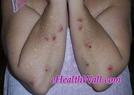
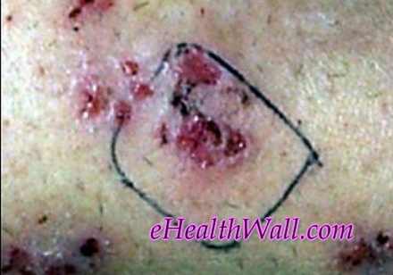
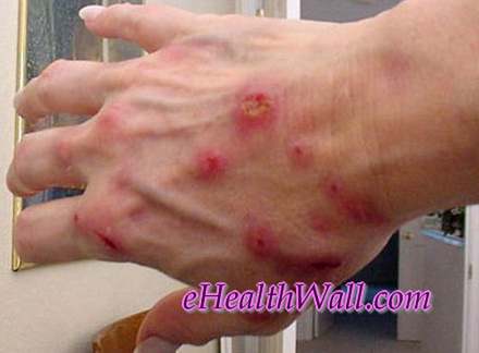
Morgellons Disease is a condition, which up to this point have not been able to have an acceptable medical condition. The name given was by an advocacy group founded in the United States of America. It started with the woman whose son suffered the symptoms that are associated with this disease. She named the condition to an ancient disease where black hairs would usually exit the skin. As up to this moment, this very rare condition is still unexplained.

Some of the health professionals claimed that this is not a medical condition that can be explained scientifically in nature but it is a condition that deals with the psychological aspect. Some believes that the symptoms being described by patients, who state that they have such kind of disease condition, are actually makings of the mind. For them, it is not true. Some are even diagnosed as having a Delusional Parasitosis. However, those who have experience having such symptoms linking to this disease, claims that it is not psychological in nature and that their symptoms are real. Some, who has this disease, are often misdiagnosed as having Lyme disease, which also manifests similar symptoms.

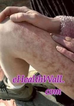
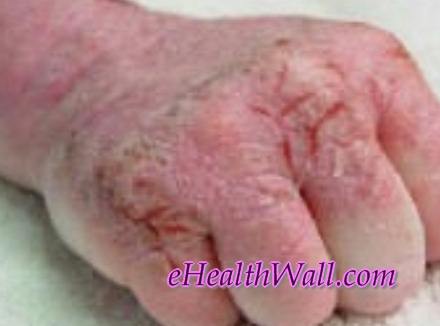
Morgellons Disease pictures (hands)

Symptoms & Signs

Persons who have the Morgellons disease say that they manifest the following common symptoms:

Physical symptoms

  1. biting, creeping and stinging sensation
  2. eruption of fibers from the lesion
  3. granules on skin
  4. black speck-like material
  5. fatigue
  6. hair loss
  7. vision loss
  8. periodontal disease

Mental symptoms

  1. Short term memory loss
  2. Thought process is impaired
  3. Attention deficit
  4. Depression
  5. Neurological problem

The persons who affirms that they have the disease describes that an insect or insects are crawling and digging a hole in their skin, making the person feel as if something is biting or even stinging them. Plus there are lesions that accompany these sensations that they are feeling. The lesions are the type that does not heal. It is correlated with structures that are unusual and bizarre that has an appearance of multi colored filamentous, fiber like strands. The color of these lesions varies from white, blue, black or red. Aside from that, their hair’s become dry and brittle.

Hence, are more prone to hair loss. Also, they experience a somewhat granular feeling in their eyes. They sometimes have problems with regarding their visions. They also experience fatigue due to total exhaustion in scratching the itchiest areas of their body where they claim to be the location of the insects that are crawling. In addition, person with this disease are reported to experience having short term memory loss, along with having an unclear way of thinking.

They find it difficult to process thoughts. Thus, they are unable to express themselves in as normal way as they can. They have ADHD or attention deficit disorders for they are unable to concentrate. And lastly, they experience depression during the actual phase of the disease.

Causes

The exact etiology of this disease condition is not yet, as of the moment, been known. The cause of this unknown skin disease has been in debates. Some says it is environmentally caused. While others suggest it is psychological in nature. Those who states that this disease is caused by environment factors leads to the findings that it is associated with three bacterial pathogens namely Borrelia species, Chlamydophila pneumonia and Babesia species. On the other hand, those who says that this disease is linked to psychological condition, believes that it is analogous to delusional parasitosis and reflects that of obsessive compulsive disorder of skin picking oneself.

Treatment

There are two treatment options that are believed to cure this disease. This includes Holistic treatment options, and medical treatment options.

Holistic Treatment Options

Since it is a skin disease, when bathing, the use of Epsom salts and the like may help in preventing the itchy feeling of the disease. Consumption of vegetables and fruits, which is washed in chemically free water, that have low sugar components are advised. Also wearing clothes only once before washing it and the use of baking soda, salt, borax and botanical blends to wash the clothes is very much recommended.

Drinking water that is only purified will help in the body purification and prevents dehydration. The usage of ammonia, bleach, pesticides, mineral oils/fragrance, and any other cleansing items that are of similar kind should not be further use for it might be the cause the disease to get worse. Holistically, the main aim is the totality of living healthily minus the chemicals.

Medical Treatment Options

The treatments that are under this are limited. It includes the same medication used to treat Lyme disease. The main reason why some of the physician misdiagnoses this condition is the fact that it can be treated with the same medication that treats other disease conditions such as Lyme disease.

Antidepressants

As mentioned earlier, one of the mental symptoms that is associated with this disease is depression. Antidepressants are prescribed in treating this symptom along with the treatment of ADHD or attention deficit disorder.

Antibiotics

Antibiotics offer relief. Antibiotics may be bacteriostatic or bactericidal in nature. Those who claimed earlier that this disease is caused by bacteria will prescribe certain antibiotics.

Anti fungal & Laser Skin Treatment

Medications such as anti fungal may be beneficial for people having this kind of disease. Laser skin treatments also offer a relief of this skin disease. Although, it might be an expensive option to some, still it provides great and effective results.

Complications

The disease itself is undergoing such further studies. The exact complications haven’t been really been exposed. However, there is a study that shows that the parasites that are associated with this disease, weakens the function of the immune system. The parasites are said to be carriers of protozoan, Lyme and or Toxoplasma Gondii.

Morgellons Disease Research

The research of this unusual disease was brought about a mother searching for answers to her sick child who manifest the symptoms associated with this kind of disease. The aim of the overall research is to gain knowledge of the pathology, etiology and medical treatment that will aid in curing the disease and understanding further the patients who has the disease per se.

As mentioned earlier, this disease is still undergoing numerous studies in order to pinpoint the exact etiology and come up with the exact treatment for this disease. Hence, persons who claim to manifest the symptoms, have to be patient enough with regards to the treatment. The physicians, due to the fact that there is a poor knowledge of this condition, would tempt to give medications in a trial and error method.

Pictures

Here are some pictures of Morgellons disease

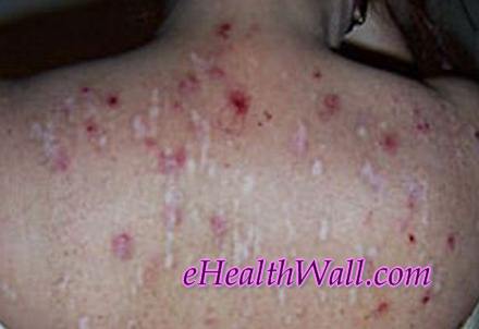
Morgellons Disease skin images

Morgellons Disease pics

Morgellons Disease thighs pictures

Morgellons Disease images

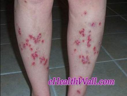
Morgellons Disease on back of legs

Morgellons Disease pics

No responses yet

Smelly Feet – Causes, Treatment and Remedies

Feb 16 2012 Published by under Diseases & Conditions

What is Smelly Feet?

Smelly feet are also called as Foot Odor is a type of body odor that affects the feet that is considered as to be unpleasant smell. The unpleasant smell that comes from the feet is normally due to sweat. Sweat particularly has no odor but it can aide in development of bacteria to grow and produce a bad smell. Having smelly feet is uncomfortable as well as embarrassing in public. It is caused by bacteria called brevibacteria that consumes the dead skin of the feet that is common on foot soles and between toes.

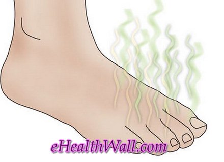
smelly feet cartoon picture

Causes

  1. Sweaty feet
  2. Poor hygiene
  3. Wearing shoes for long hours because of lack of air ventilation
  4. Too much hair on the feet can also contribute to odor intensity by adding surface area to the bacterial growth
  5. Shoes and socks that is made of synthetic materials that will lead the feet to sweat more
  6. Fabrics like nylon can lead to sweat and odor because they cannot absorb the sweat effectively
  7. Some foods can make the body smellier. Foods like garlic, curries, chili, onions and alcohol.

Treatment & Prevention

  1. Maintain good hygiene is a first step for preventing foot odor.
  2. Keep your feet clean by washing your feet regularly and used anti-bacterial soaps that is good to get rid and kill bacteria causing foot odor.
  3. Socks should be change regularly.
  4. Cotton Shoes and Socks or those with natural fabrics that can absorbs sweat .
  5. Using of Antiperspirant or Deodorant products for the feet. Antiperspirant products works by inhibiting the release of sweat in the body. Deodorants do not decrease sweat but they prevent odor that is cause by a bacteria.
  6. The feet should be kept dry all the time. Have a shoes and socks that can absorb sweat and that can help in passage of air to maintain the feet dry.
  7. Wear shoes that is completely dry because it will help to reduce bacteria causing the foot odor
  8. Use of Cedarsoles that can eliminate foot odor. Cedarwood is known for antifungal properties and it can work on the feet.

Home Remedies

  1. Apply tea to the foot : Using a soak of warm water and tea and soak your feet for at least 30 minutes and it will help eliminate the problems of smelly feet
  2. Use of Alum that can be seen at the spice section of the supermarket. Mix 1 teaspoon to a 1 gallon of water and soak your feet for at least 30 minutes.
  3. Use of Apple Cider Vinegar to control the odor. Mix one-third of the apple cider vinegar to a small pan of water or a pan that you can soak your feet and do it several times in a week.
  4. Use a baking soda and put it in your shoes because it will help to absorb odor as well as moisture.
  5. Use of Anti-bacterial soaps to kill and get rid the bacteria causing foot odor.
  6. Using isopropyl alcohol for at least two weeks. It’s cheap and highly effective cure for foot odor.
  7. Sprinkle some cornstarch or baby powder on your shoes every time you will wear it. This will help to absorb moisture from sweat and also it helps to keep your feet dry.

No responses yet

Flesh Eating Disease – Pictures, Symptoms and Treatment

Feb 14 2012 Published by under Diseases & Conditions

What is Flesh Eating Disease?

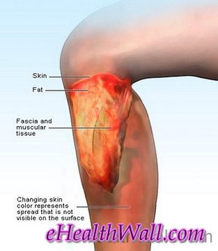
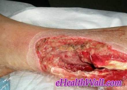
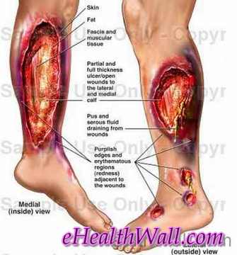
Flesh eating disease is also known as necrotizing fasciitis, which means decaying skin. It is a rapid progressing type of bacterial infection that destroys the skin, fat and tissue that covers the muscle in a short time span, within 12 to 24 hours. Actually, the flesh is not eaten but the skin is dying as a result of the infection that the person has.

The bacteria, associated with this disease, cuts off the blood supply. Hence, the tissues die and go through the process of decay, medically termed as necrosis. It is caused by a bacterium; group A beta hemolytic streptococci which is common culprit in this type of disease, which targets the layers of tissue, which is termed medically as fascia, in your muscles. It is a serious and very rare disease condition. In occurs in 1 out 4 people.

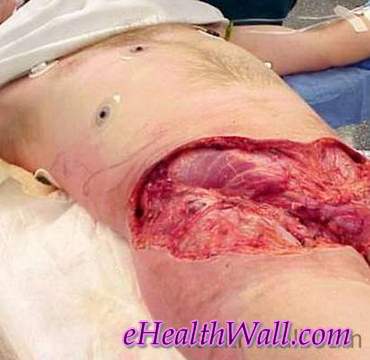
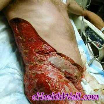
Flesh Eating Disease pictures

Picture 1 – Flesh eating disease at groin

How do you get Flesh Eating Disease?

There are many different ways in acquiring the bacteria which causes the disease condition. The one common reason is when the culprit bacteria enters your body through an open wound, from a burn, a cut, surgery, or an insect bite. You can also get it:

  1. When one has surgery sites or in tumors or in gunshot wounds
  2. when one has muscle strain or bruise
  3. Wounds coming in contact with saltwater fish, oyster which are both raw and ocean water or when you are handling animals from the sea like crabs.
  4. When one has a weak immune system
  5. When one is suffering from chronic disease in the heart, lungs, or liver disease.
  6. When one has recent contact with someone who is positive with the disease condition.
  7. When one has chickenpox, all though not all having chickenpox ends up with this disease condition.

It is important that you keep in mind that this flesh eating disease occurs very rarely. So even if you have the factors mentioned above, the chances of acquiring it is low.

What causes Flesh Eating Disease?

The necrotizing fasciitis is caused by many bacteria. Some of these bacteria are proven to cause infections like impetigo or strep throat. This kind of bacteria causes mild form of infections. In rare case, it can result to a much more dangerous infection

Is Flesh Eating Disease Contagious?

This disease, which is somewhat contagious, can be passed from one person, who has an immune system that is low, to another by mere close contact, through kissing, touching the infected wound, or when an infected person cough or sneeze, with the one who has the bacteria. The patient having the disease condition is not likely to be contagious and the transmission points are through objects that are inanimate in nature.

Flesh Eating Disease Symptoms & Signs

Early recognition of the symptoms associated with this disease includes the following:

  • Altered Mental State. Toxic effects of these bacteria can result to disorientation, confusion or mental dullness. When the bacteria have spread to the bloodstream, shock can develop and in the long run can lead to coma.
  • Pain. An early sign of this disease condition is severe pain. This is due to the fact that the nerves in the affected area are dying; hence there is poor circulation of blood, leading to pain.
  • Heart Palpitations. Infection and High Fever leads to increase heart rate, which is leading to heart palpitations.
  • Local Skin Warmth. The affected area appears, initially, red and is hot to touch. The inflammatory process is triggered. You must take note that the skin changes from red to purple to dusky gray.
  • Swelling. The area infected is swollen plus the skin is shiny and taut. It usually takes place within 30 minutes.
  • Blisters. A popping and crackling sound is produced when you press the skin, blisters formation may accompany it.
  • Foul Odor. When the skin and soft tissues go through the dying stage, it will eventually produce a foul odor that emanates from the wound.
  • Ulceration. There is an opening of the skin which is developing when the infected tissue of the skin dies.
  • Malaise. Extreme fatigue along with headache, nausea and generalized weakness is another symptom if this disease condition.
  • High Fever. Chills and sweating are common along with high fever which runs through 38.9 to 40.6 degrees Celsius or 102 to 105 degrees Fahrenheit.
  • Dizziness. The disease causes blood pressure that is low which results to the feeling of fainting or being dizzy.

Flesh Eating Disease Treatment

Once there is a confirmed diagnosis, steps must be taken as soon as possible. When the treatment is not done or undertaken, this disease condition can be fatal.

Surgery

The primary step is debridement of the infected area which is done surgically.

Medication – Antibiotics

In addition, antibiotics are given intravenously which should be administered as soon as suspicion arises.

Hyperbaric Oxygen Therapy

This helps in prevention of tissue necrosis or death and aids in the promotion or recuperation.

Early treatment is necessary. The sooner the infected area is treated, the more likely you will avoid complications that are serious and will recuperate from the bacterial infection.

Flesh Eating Disease Prevention

There are two important things you have to remember to prevent acquiring this disease condition:

Seek medical attention. If you suspect someone or manifest the symptoms that have been mentioned above. You can take antibiotics also as a precautionary action.

You should take good care of any minor cuts or wounds. Wash the infected area with soapy warm water, keep it always dry and clean and always cover it with bandage.

Flesh Eating Disease Pictures

Here are few pictures of flesh eating disease

Flesh Eating Disease pictures

Flesh Eating Disease images

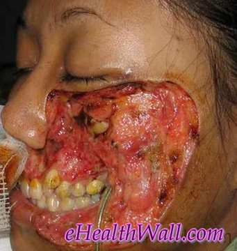
Flesh Eating Disease on face images

Picture 2 – Flesh eating disease on face

Flesh Eating Disease pics

Flesh Eating Disease (necrotising fascitis)

Picture 6 – Flesh eating disease on right side of chest, abdomen & thigh

No responses yet

Microcephaly – Pictures, Definition, Causes, Life Expectancy, Prognosis

Feb 10 2012 Published by under Diseases & Conditions

What is Microcephaly?

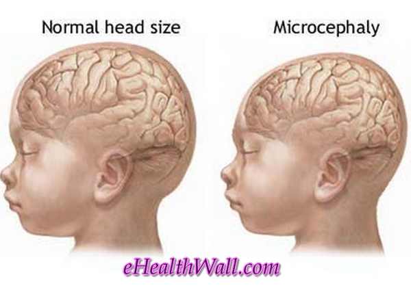
Microcephaly is a neurodevelopmental disorder where in the person’s head is small for an average person’s gender and age. It is a birth defect of which children are born with a smaller head. Since their head is small, their learning development is also affected. It interferes with the cerebral cortex’s growth.

Microcephaly is a medical definition that literally means “small head”. It has been associated with mental retardation but not all children having small head are known to have mental retardation.

Microcephaly with normal head comparison

Picture 1 – Comparison of normal head with microcephaly

Image source – Adam inc.

Microcephaly Types

Primary Microcephaly

This is a kind of microcephaly. It is sometimes known as a true microcephaly or Microcephaly Vera. It is an inherited disease condition that is due to genetic mutations. There are four mutations that lead to the cause of this disease condition, this genes are: MCPH1, CK5RAP2, ASPM, and CENPJ. Genetic mutations in primary microcephaly debase the development and brain growth because this plays an important role in brain cell division.

Congenital Microcephaly

When a person has congenital microcephaly, it means that the person has this kind of condition at birth. Studies show that before a person has a diagnosis of congenital microcephaly, it may follow intrauterine infections like toxoplasmosis, rubella, and cytomegalovirus.

Microcephaly Causes

The cause of microcephaly may be due to the ingested substances which are considered harmful. These kinds of substances which mothers take during fetal development may lead to their children having a diagnosis of microcephaly. It may also be associated with genetic syndromes.

According to theoretical studies conducted, the following predisposing factor may affect the fetus’ normal head development during the pregnancy state:

  • Metylmercury poisoning – It will affect the fetal development especially during the pregnancy state.
  • Cytomegalovirus,  varicella infection or rubella – These are the infections of the fetus during the pregnancy state.
  • Lack of proper nutrients and vitamins in the diet – It may lead to this diagnosis since when you lack nutrition especially during pregnancy will really affect the development of the fetus.
  • Exposure to harmful substances or chemicals – It will lead to the risk for abnormalities in the brain
  • Untreated phenylketonuria – It is birth defects that compel the ability of the body to breakdown the phenylalanine which is an amino acid.
  • Prescription of illegal drugs – It will lead to abnormalities in the brain development.
  • Alcohol consumption during pregnancy – This will lead to the risk of abnormalities in the brain.
  • Cerebral Anoxia – It means decrease oxygen to the fetal brain will lead to complication of pregnancy and impairment of oxygen delivery to the brain of the fetus.
  • Craniosynostosis – It is the premature fusing of the joints which may lead to microcephaly.
  • Chromosomal abnormalities – Such abnormalities like down syndromes may lead to microcephaly.

Microcephaly Diagnosis

This disorder which is congenital can be diagnosed before birth through the use of prenatal ultrasound. It is a diagnostic imaging technique that uses sound waves that are high frequency.  Other confirmatory diagnostic tests that the physician can do are the following:

  1. Urine Test
  2. Blood Test
  3. MRI or Magnetic Resonance Imaging
  4. CT scan or Computed Tomography Scan
  5. X-ray
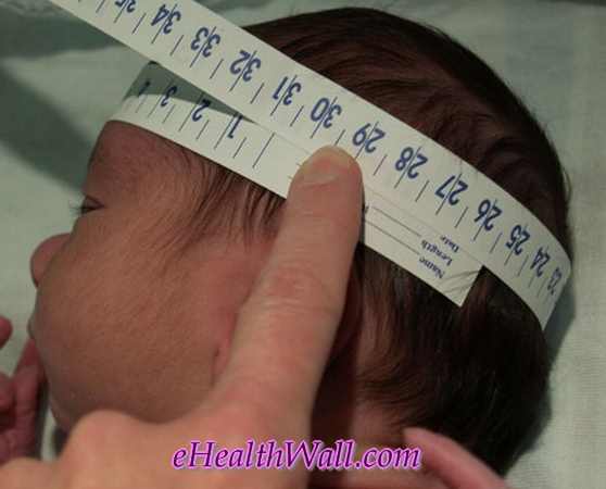
  6. Head Circumference
head circumferenec measurement for microcephaly diagnosis
Picture 2 – Head circumferenec measurement to diagnose microcephaly
Image source – newborns.stanford.edu

Microcephaly Treatment

Up to this point there is no specific treatment that will resolve the microcephaly condition. The treatment that is discussed in this article will focus on decreasing the impact of the neurological disabilities and associated deformities.

  • Therapies such as speech, occupational, and physical therapy aid in minimizing the dysfunction and maximizing their potential abilities.
  • Other physicians will give prescribed medications to control hypersensitivity, neuromuscular symptoms and seizures. The treatment is a supportive kind of treatment.
  • You can also have genetic counseling to let the families know the risk for microcephaly during the next pregnancies.
  • The medical team working in the child diagnosed with microcephaly will be the one who will give the educational information and guide them into the improvement of the child’s well being.
  • Microcephaly is a lifelong condition which is non-correctable. There is a positive reinforcement to help in encouraging the child and to boost his or her self-esteem and encourage independence.

Prognosis

Its prognosis varies and will depend on the medical condition of the patients.

  • While some children may have minor disability, some will grow and develop normally and will have normal intelligence. It really depends on the patient himself or herself. The prognosis of a person diagnosed with microcephaly is really not known.
  • According to reports, most children died within his or her first year. Others will say that the person diagnosed with microcephaly will have a varying prognosis.
  • Its prognosis will usually depend on the symptoms that he or she is experiencing. In most case, it was reported that most patients will survive and will meet their developmental milestones normally.
  • Yet, some patients diagnosed with this disease condition will be able to experience delay on their developmental milestones and will really need professional help and support.
  • The person who usually supports them, aside from their family members, are their therapist. In determining the exact prognosis that a patient has with regards to being diagnosed with such disease condition, is the need for them to undergo evaluation from their trusted and specialized physicians and they must do such as soon as possible.
  • As mentioned earlier, the prognosis will depend on the condition of the patient himself or herself. The microcephaly is not really evident after birth, but it is revealed as the person, or the child for that matter, develops and grows.

Life Expectancy

The general life expectancy of the children diagnosed with microcephaly is reduced. The prognosis and the life expectancy work hand in hand. If for instance, the child has a poor prognosis, the life expectancy for that child is low. Yet some experts will say that the life expectancy with a person diagnosed with microcephaly will usually last for 20 to 35 years old, if they are lucky enough. With the right support mechanisms from family members, loved ones, and physicians, the life expectancy will be high and vice versa.

Pictures

Here are some pictures of microcephaly (small head).

Microcephaly images

Microcephaly (small head) pictures

Microcephaly pictures

Microcephaly pics

No responses yet

Next »

© 2017 eHealthWall.com. All Rights Reserved. Privacy Policy
This website is for informational purposes only and Is not a substitute for medical advice, diagnosis or treatment.