Archive for May, 2012

Coulrophobia – Definition, Facts, Treatment, Symptoms, Causes

May 31 2012 Published by under Diseases & Conditions

Coulrophobia Definition

All of us have fears or phobias, which are defined as persistent and irrational fear. When you have phobias of clowns, you have the phobia called Coulrophobia, which is a Greek word which was culled out from the Greek word kolobathristes . Symptoms of coulrophobia is like the one who walks in stilts. Stilts were the name given to clowns during those times.

This kind of phobia is commonly reported in children but adults as well as teenagers can have phobias such as this. The persons who have phobias in clowns usually acquire it because they have a bad experience with such in a personal situation. Sometimes, clowns are used as the antagonist in some movies. It is also defined as an extension of fear of having to cover one owns face with the use of paint. The axiom of hiding features which are recognizable under the facial layer by using paint which can also bring about Coulrophobia.

coulrophobia

Coulrophobia Image

Coulrophobia Symptoms

Despite the fact that clowns are known to bring fun or give way a humor to their audience, persons who suffer from the fear of the clowns or those who have Coulrophobia will experience panic attacks when faced to faced or upon the encounter of clowns. Other associated symptoms include:

  • Anxiety
  • Feeling of dread or terror
  • Palpitation or irregular or rapid heartbeat
  • Having shortness of breath
  • Intense perspiration
  • Nauseated
  • Mild discomfort
  • Loss of mental stability and act as if one is going insane
  • Fainting
  • Lightheadedness
  • Stomach pain
  • Sweaty palms
  • Feeling of terror
  • Mouth becoming dry
  • Limbs will start to shake
  • Paranoid thoughts run around whenever one encounters clowns
  • Unable to speak normally

Coulrophobia Causes

Negative experiences

Apparently, there are no medical or scientific inclinations to why such persons experiences Coulrophobia. Persons who have Coulrophobia make it a point that they avoid any circumstances which may lead them to experience meeting up with clowns. They try to avoid any kind of clowns or escape from the encounter of their biggest fear, which are clowns. The one typical thing why person experiences Coulrophobia is because they have encountered an experience with clowns, which they considered as traumatic for them. Aside from the negative experience, clowns are also used, as mentioned earlier, in horror movies which brings about another not so positive experience with clowns.

When clowns who are seen on television becomes huge persons in reality

Some children who are fond of children’s characters, which are fantasy such as clowns and Santa Claus, sometimes becomes terrified when what they see on television seems to huge or tall when they come face to face with such in the real world. Hence, they develop a sense of Coulrophobia.

Antagonist portrayal of clowns

Others persons, grow a fear of clowns upon the viewing of movies that have clowns portraying a monster or murder role. There are such movie makers which likes the irony of persons who brings happiness to others as someone who instantly becomes a painful source. Hence, they use clowns to portray the boogeyman, killer, and many other antagonist roles which may lead to persons having fear of clowns.

Learned behavior of Coulrophobia

Studies have shown that children who have parents having Coulrophobia also grown up to have one. There is a theory that phobias can be a learned behavior that we may grow up with because our surroundings tell us to be feared of something. Hence, we grow up fearing of those things.

Coulrophobia Facts

Clowns are funny performers who wear wigs, colorful make ups, and costumes. Its job is to entertain and provide the audience happiness. However, in the case of persons who have Coulrophobia what they offer are panic attacks. Statistics shows that one out of seven persons has Coulrophobia or has a fear of clowns. Hence, making this kind of phobia is very rare in occurrence. The persons with Coulrophobia do not only fear the clowns who they see with their own eyes, they also have encounter panic attacks whenever someone says the word clowns. Persons with Coulrophobia should not be judge accordingly but they have to be understood and accepted.

Coulrophobia Treatment

Gradual Exposure Therapy

The treatment suggests to persons who have phobias on clowns is through gradual exposure therapy. It is what psychologist would recommend, which is to face your fears for avoiding it will not help in any way. It may be easy to say such phrase but you can be accustomed to it in the long run and by the time you know it, you will be able to conquer your fear of clowns. Through doing such therapy you become more at home or familiar with the clowns. So how do you start, you might ask. The first thing that you do is to show them either pictures or videos that has clowns which displays a friendly clown and not show them horror films that depicts clowns as a monster or portraying a scary role. The goal is to find comfort or be comfortable with clowns. In such a way, the person who has Coulrophobia will be desentisized with their fear.

coulrophobia Images

Gradual Exposure Therapy for Coulrophobia

Relaxation therapy

The person who has Coulrophobia is suggested to engage in therapies that relaxes them such as yoga, meditation, as well as acupuncture. This will not only help them deal with their fears, this will also bring about good health.

Free association therapy

Persons who have been dealing with Coulrophobia are suggested to consult a psychotherapist and recommended to undergo session that they call as free association therapy. During this therapy, the person is called to say whatever that comes into his or her mind when exposed with a certain thing. The person undergoing this therapy must be comfortable in saying things that are on his mind, there should be no instances that he or she should be embarrassed by saying such. This is an effective method that allows the person to voice out his or her negative experience and be able to understand what the person is going through.
No responses yet

Onychocryptosis – Treatment, Symptoms, Causes, Prevention

May 29 2012 Published by under Diseases & Conditions

What is Onychocryptosis?

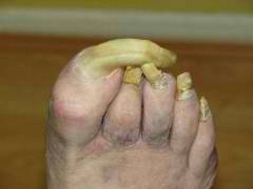
Onychocryptosis is a medical terminology which means ingrown toenails. It is a condition which can affect various kinds of persons. Another term for Onychocryptosis is unguis incarnatus which can be located at the toenails and fingernails.

Persons with Onychocryptosis  causes an excess in the lateral part of the curvature of the person’s nails and traps the epithelial cells which is found in the pinch and sulcus areas which is located in the nailbed.

It occurs when the person’s nail’s wall and edge becomes pushed up which results to the occurrence wherein the nail cut into the person’s nail wall. Other experts would say that there is an abnormal extension of the person’s toenail which eventually results to such nail disorder.

This becomes a very painful experience to a person having Onychocryptosis. Some persons having this kind of nail disorder will encounter repeated incidences of infection as well as pain. It may become also a chronic kind of nail disorder. The nail disorder is reportedly affects the big toe’s outer edge.

Onychocryptosis

Onychocryptosis Image

Onychocryptosis Symptoms

The symptoms that a person with Onychocryptosis may able to encounter the following:

  • Painful to touch
  • Irritation
  • Swelling
  • Redness
  • Sensation of warmth
  • Pus (with or without)
  • Watery discharge
  • With blood or without blood
  • Presence of infection
  • Fever

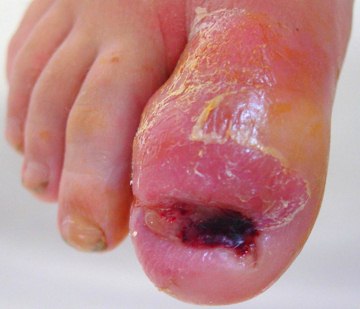
Onychocryptosis

Ingrown nail infection

Image source: img.medscape.com

Onychocryptosis Causes

Persons with Onychocryptosis will be experience the nail disorder because of the following etiological factors such as:

  • Wearing of shoes that are ill-fitting
  • Trimming of the person’s toenails done improperly
  • Trauma or Injury of the person’s nail like nail peeled off or ripped nail which leads to ingrown toenail
  • Fungal or yeast infections
  • Having a bad habitual nail care
  • Softening of the plates of the nails
  • Prescribed medication’s side effects such medications like acitretin and isotretinoin
  • Socks or hose which are too tight
  • Toenail deformities such as having excessive thicker nail plate

Onychocryptosis Treatment

With regards to treating Onychocryptosis, the person can either do the following:

Home Remedies

  • Massaging gently the inflamed skin
  • Soaking the person’s affected foot with water which is warm. Do this 3-4 times daily. Also keep the person’s affected toe dry after. This is otherwise known as warm water soaks therapy
  • Placing a small dental floss or cotton under the person’s nail otherwise known as a cotton-wick insertion therapy
  • In mild case of Onychocryptosis, the patient is suggested to dip the affected toes into a water which warm and which has a mixture of Epsom salt
  • Trim your nails, which should be done straight across the top
  • Washing the affected foot with soap and water and do this at least two times a day
  • Using glutter splinter through the use of slit plastic tubing which will aid in keeping the lateral nail folds and the nail apart
  • Bandaging the affected toe

Pharmacological Medications

  • Applying mild antiseptic solution as prescribed to the affected toe
  • Topical or oral antibiotics as prescribed which will prevent infection and aid in the fast recovery of the affected toe
  • Pain relievers which are prescribed to provide pain relief
  • Iodine application as prescribed

Surgical Procedure

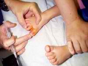
Surgical removal of the ingrown toenail as suggested and done by an expert physician, that is especially done when the condition doesn’t anymore respond to the treatments mentioned earlier. Examples of surgical procedure done are partial nail removal or total matrixectomy which are done with severe Onychocryptosis.

In the successful treatment of Onychocryptosis, you must work hand in hand with the podiatrist or dermatologist or perhaps your trusted family doctor. They will help you suggest and prescribe treatment which will be beneficial for you to be able to proceed to the recuperation stage.

total matrixectomy picture

Total Matrixectomy to remove Onychocryptosis

Onychocryptosis Prevention

In preventing the nail disorder called Onychocryptosis, it is advised that you should do the following things:

  • When trimming, be sure that you will use a sharp and clean nail trimmer and never use scissors in trimming your toenails
  • Never tear or pick at your nails
  • You should take it to a habit that you will trim your toenails straight across the top
  • With regards to shoes, you should wear shoes that fit your toes properly well. Your shoes should have space around your toes
  • Keep your feet dry and clean always as much as possible
  • When you are diabetic, have visual or vascular problems, it is advisable that you go on routine examination of the foot and nails to prevent Onychocryptosis
  • In trimming your nails, you must avoid overcutting or curving your toenails too short especially at the edges part
  • As much as possible, avoid doing physical activities which involves stopping suddenly like basketballs which have the tendency to jam the toes in the front of the person’s shoes
  • It is also advisable that you wear loose socks
  • Avoid repeated toenail injury
  • Maintaining a good hygiene for your foot
  • You should also avoid dropping things in the toenail

By doing such, you are able to avoid experiencing Onychocryptosis or ingrown toe nails.

No responses yet

Degenerative Arthritis – Treatment, Symptoms, Causes, Types

May 26 2012 Published by under Diseases & Conditions

What is Degenerative Arthritis?

Degenerative arthritis is a medical condition involving the wearing or degeneration of the cartilage in the joints leading to contact between the two bones. Degenerative arthritis is also termed as osteoarthritis. The cartilage in the joints is the one responsible for the motion of the joint as well as limiting the friction and tension in between the bones in the joints.

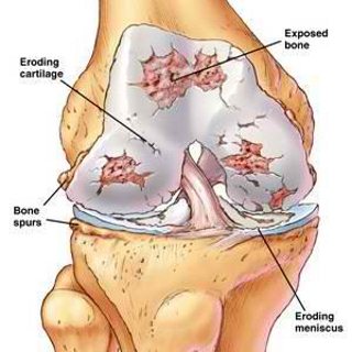
In degenerative arthritis, the cartilage is damaged leading to the increased in the friction between the bones. Degenerative arthritis is most commonly seen in weight bearing joints in the body such as the neck, knees and hips, but it can also occur in other joints. The damage in the joint also causes certain changes in the joint capsule as a result of inflammatory response and healing.

The initial response of the joints to damage is the formation of osteophytes in the damaged cartilage. Because of this, there is further narrowing in the joint space. The osteophytes result in the growth of subchondral cysts in the cartilage.

Degenerative Arthritis Image

Degenerative Arthritis Image

Image source: health-reply.com

Symptoms of Degenerative Arthritis

Symptoms of degenerative arthritis include:

  • Joint pain – The constant friction in the bones in the joint causes inflammatory response on the area causing pain. Pain is characterized as burning and sharp on the joint area, including the muscles and the joints. Cold temperatures usually aggravate the pain. The pain is relieved by gentle motions, but becomes severe with high impact use.
  • Stiffness of joints – The other structures such as the muscles, ligaments and tendons in the area are also affected leading to limited motion of the joint. The cartilage that normally allows for smooth motion of the joints is damaged that causes stiffness in the area.
  • Swelling of the joint – The joint also appears swollen because of underlying inflammatory process in the area.
  • Crepitus – When moving the joint, a characteristic crackling sound is heard because of contact between the bones in the joint.
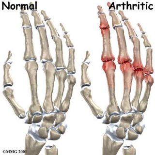
  • Heberden’s nodes – Heberden’s nodes are bony enlargements on the distal interphalangeal joint. These appear because of the growth of subchondral cysts in the joints. These are often not painful.
  • Bouchard’s nodes – These are similar to Bouchard’s nodes only that they occur on the proximal interphalangeal joints.
  • Joint effusion – The joints may also develop collection of water or fluid as a result of accumulation of synovial fluid in the area.

Degenerative Joint Inflammation  Image

Swelling of the joints

Degenerative Arthritis at Various Places

Degenerative arthritis is most commonly located at weight bearing joints, but it can also occur in all joints in the body. The most common locations include:

Degenerative Arthritis of the Spine

The spine is a common site of degeneration because of bearing the weight of the back and the head. The spine can also be affected by trauma and various inflammatory conditions such as ankylosing spondylitis leading to degenerative arthritis.

Degenerative Arthritis of the Knee

The knee is also a common location of the condition especially among obese patients because of increased tension in the area as the knees bear the weight of the body.

Degenerative Arthritis of the Neck

The neck or cervical spine carries the weight of the cranium and its content. It also has several range of motion, which increases the stress in the cervical joints.

Types of Degenerative Arthritis

Primary

Primary degenerative arthritis is a type of joint disorder that is caused by the actual destruction of the joint caused genetic factors. Primary osteoarthritis causes the water content of the cartilage to decrease. The loss of the fluid is associated with aging. The absence of protective fluid or proteoglycan causes the cartilage to become susceptible to damage. Primary osteoarthritis is classified into nodal osteoarthritis and erosive osteoarthritis. Nodal osteoarthritis involves the formation of nodes in the joints, while erosive type involves the progressive destruction of the joint. Erosive from tends to be more severe, but less common than nodal osteoarthritis.

Secondary Osteoarthritis

Secondary osteoarthritis is a type of arthritis that results from the degeneration of the joints as a result of underlying conditions that hastens the degeneration of the weight bearing joints.

Causes of Degenerative Arthritis

  • Degenerative arthritis is caused by certain factors. Primary and secondary osteoarthritis have different causative factors. The main cause of primary osteoarthritis is genetics. Aging is an aggravating factor because of the normal degeneration of the joint. Secondary osteoarthritis is caused by various factors such as:
  • Obesity – Obesity is one of the most common causes of secondary arthritis because of increased tension on the cartilage on the weight bearing joints.
  • Congenital disorders – Congenital disorders of the joints involve the presence of defects on the area since birth leading to increased risk for developing degenerative arthritis.
  • Inflammatory disorders – Inflammatory disorders such as Lyme disease and Perthe’s disease increases the tendency to develop similar inflammatory condition in the joint.
  • Metabolic disorders – Disease such as diabetes and Wilson’s disease also leads to increased degradation of the joints.
  • Trauma – Trauma to the joints or ligaments also leads to tearing and injury to the area.

Treatment of Degenerative Arthritis

Various treatments for degenerative arthritis include:

  • Weight Loss – Losing weight is an important management for osteoarthritis to prevent further stress and tension in the joints.
  • Exercise – Exercise is also essential for patients with osteoarthritis. Stretching exercises are more beneficial because it strengthens the muscles and structures around the joints thereby providing more support.
  • Administration of Anti-inflammatory medications – Medications such as ibuprofen and naproxen relieve inflammation and pain. Aspirin may also be used, but it can cause adverse reactions such as gastric irritation and bleeding on the joints. Newer medications such as celecoxib can prevent these effects. Cymbalta, an antidepressant drug is also effective in reducing inflammation and pain.
  • Apply warm compress – Warm compress may be placed for 15 minutes three times a day to relax the muscles in the area relieving pain.
  • Alternative medications – Chondroitin and glucosamine can also be given to patients as food supplements. These substances are a natural component of the synovial fluid. Taking these drugs may help in the synthesis of collagen in the cartilage thereby increasing the integrity of joints.
  • Surgery – Surgery becomes the last treatment for degenerative arthritis. This may involve osteomy or the removal of the subchondral cysts and chondroplasty or the repair of the cartilage. Severe cases may require joint replacement by artificial prostheses.

No responses yet

Myxedema Coma – Treatment, Symptoms, Causes, Diagnosis

May 22 2012 Published by under Diseases & Conditions

What is Myxedema Coma?

Myxedema coma is a condition resulting from decompensated or unmanaged hypothyroidism. Hypothyroidism is the condition wherein the thyroid gland does not secrete adequate amount of thyroid hormones leading to problems in various metabolic processes in the body.

Myxedema coma present as a normal hypothyroidism based on laboratory tests, but other serious conditions can trigger the development of the state. The condition results from severe and persistent low thyroid hormones that cause loss of brain function. Myxedema is the most severe form of hypothyroidism and may result in death.

Myxedema coma is also considered a rare development of hypothyroidism. It is seen more in women and elderly patients with hypothyroidism. Myxedema coma is also increased during winter months because of increased cold intolerance experienced by patients.

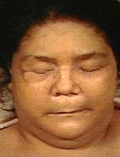
Myxedema Coma Image

Myxedema Coma

Myxedema Coma Symptoms

Symptoms of myxedema coma result from the affectation of the brain as well as other organs in the body. Generally, the symptoms include slowing down of the functioning of the body. These include:

  • Altered mental status – Patients often exhibit alteration in the level of consciousness that may start from disorientation to lethargy and eventually coma.
  • Seizures – Seizures are a common manifestation of myxedema coma because of altered firing of nerve impulses in the brain.
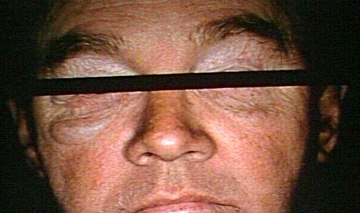
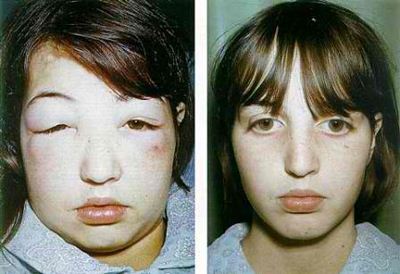
  • Anasarca or generalized edema – Poor metabolism of fats, proteins and carbohydrates leads to reduction in the osmotic pressure in the blood. The fluids tend to shift from the intravascular space into the interstitial space. Edema is often seen on the periorbital area and the tongue. Severe edema may involve the lungs and the heart membranes.Myxedema Coma Image

Anasarca as a symptom of Myxedema Coma

  • Hypothermia – The core temperature of the body is often reduced up to 26.6 degrees Celsius. Patients initially have cold intolerance that progresses to severe loss of heat from the body.
  • Hypotension – The cardiovascular integrity is also affected leading to a loss of vascular tone as a result of impaired heart contractility as well as loss of fluid in the blood vessels.
  • Hypoglycemia – The reduction on the metabolic rate affects the conversion of carbohydrates into glucose leading to hypoglycemia.
  • Bradycardia – The reduced metabolic rate also slows down the contraction of the heart muscles. Patients may suffer from cardiac arrest when not treated promptly.
  • Hyponatremia – The increase in fluids in the body dilutes the blood and leads to hyponatremia or the low level of sodium.
  • Hypoventilation – The respiratory system is also affected due to central nervous system depression.

Myxedema Coma Causes

Myxedema coma initially starts as hypothyroidism. Hypothyroidism may be caused by several factors such as radioactive exposure of the thyroid glands, overtreatment of hyperthyroidism, thyroid surgery or hypothalamic and pituitary problems, which leads to poor stimulation of the thyroid gland to produce hormones.

Myxedema coma is usually triggered by several factors, which increases the burden to the thyroid gland to produce more thyroid hormones. These include:

  • Medications – Medication intake including anesthesia, narcotics, sedatives, lithium and amiodarone can trigger myxedema coma
  • Infections – Infections increase the metabolic rate of the body, which the thyroid is unable to support.
  • Trauma – Trauma also increases the demand for thyroid hormones in order to hasten healing.
  • Hypothermia – The presence of a very low body temperature leads to severe cold intolerance that may affect the brain and the heart cells.
  • Stroke – Stroke impedes the blood flow to the brain and may limit the functioning of the pituitary gland leading to further inactivation of the thyroid gland.
  • Hypoglycemia – Hypoglycemia is also a trigger for myxedema coma because of poor oxygenation of the brain as a result of low blood sugar levels.
  • Gastrointestinal hemorrhage – Hemorrhage may also lead to myxedema coma as a result of hypovolemia.

Myxedema Coma Pathophysiology

Myxedema coma usually develops when the patient’s homeostatic mechanisms are impaired as a result of various factors discussed earlier. The lack of equilibrium in the body intensifies the effect of hypothyroidism leading to brain affectation.

Myxedema Coma Diagnosis

The diagnosis of myxedema coma is made by testing for thyroid hormones and observing the symptoms. The presence of low thyroid hormones such as T3, T4 as well as TSH levels with the presence of the above symptoms are indicative of the condition. Other tests may be done such as blood tests and chest X-rays to determine sodium levels as well as edema of the lung and heart membranes. CPK levels are also checked using blood tests to determine altered permeability of the muscles.

Myxedema Coma Treatment

Treatment of myxedema coma usually involves emergency measures to reverse the condition and prevent death. Myxedema coma is highly fatal and patients usually die from cardiac and respiratory arrest. Patients are often placed in the intensive care unit for continuous monitoring and treatment. The first 48 hours is very critical in the treatment of myxedema coma in order to reduce mortality. Treatments include:

Intravenous Administration of Thyroid Hormones

The principal reason for myxedema coma is the reduction in thyroid hormone levels. In this regard, intravenous levothyroxine is usually employed to regain normal levels of thyroid hormones as fast as possible. Intravenous route is the choice because the gastrointestinal system is often compromised.

Antibiotic Therapy

Myxedema coma is usually preceded by an infection, thereby; bacterial etiology should be identified in order to give the most appropriate antibiotic treatment. Infections that usually result in myxedema coma is pneumonia and urosepsis.

Corticosteroids

Corticosteroids are usually administered to reduce cerebral edema as well as correct adrenal insufficiency because of hypopituitarism. Cortisol levels are initially taken to determine the level of glucocorticoids in the body. Hydrocortisone is given 5 to 10 mg per hour.

Respiratory Support

Mechanical ventilation is usually employed for patients with respiratory compromise (the presence of hypercapnia, hypoventilation and respiratory acidosis).

Rewarming

Patients also need passive rewarming with the use of warm environment and blankets. Rapid rewarding is contraindicated because of rebound effects.

Intravenous Infusion with Saline Solution

Hyponatremia requires infusion of saline to regain normal sodium levels and water regulation in the body. Intravenous infusions also correct hypovolemia.

Intravenous Glucose Administration

Glucose must also be administered to improve oxygenation of the brain cells as well as regain normal energy production.

These treatments should be given as soon as possible to prevent death. The prognosis is often undefined because of small number of incidences.

No responses yet

Plagiocephaly – Treatment, Positional, Helmet, Symptoms, Pictures

May 18 2012 Published by under Diseases & Conditions

What is Plagiocephaly?

Plagiocephaly is a structural deformity of the skull characterized by asymmetry of the head. The term specifically describes a flattening of the head to one side or at the back and sometimes associated with facial asymmetry. It is also termed as a “flat head syndrome”.

Plagiocephaly can be present at birth or develops during the early months of life. The most common form of plagiocephaly nowadays is flattening at the back because of sleeping patterns as a result of repeated pressure in a particular side or part.

what causes flat head syndrome?

Plagiocephaly should not be seen as an abnormal condition because it is normal for a newborn’s head to have flattening because of the soft skull. Plagiocephaly at the time of birth may be resolved in two months. However, the baby’s head may continuously be flattened because of external factors such as positioning during sleep.

Flattening of the head is very common among infants and should not cause alarm to parents because this does not mean any medical implications.

Plagiocephaly occurs because the sutures in the baby’s skull have not fused yet. The skull remains to be malleable and soft. Premature infants are more prone to plagiocephaly because they have softer skulls than term newborns and they are confined to the hospital longer for further management.

Plagiocephaly is different from craniosynostosis, which is a more serious skull malformation. Craniosynosotosis results from premature fusing of the sutures in the skull which makes the head deformed. Craniosynostosis should be ruled out because it requires more extensive management.

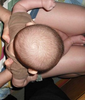
Plagiocephaly picture

Plagiocephaly Image

Plagiocephaly Types

Plagiocephaly has several types depending on the location of the flattening. These include:

1. Positional Plagiocephaly

Positional or deformational plagiocephaly is the flattening of the skull as a result of positioning the baby’s head to one side only. This may occur due to crowding inside the uterus that results to flattening or letting the infant lie in the same position for prolonged periods.

Positional plagiocephaly may be present as a flattening of the occipital or frontal areas of the skull and may cause facial asymmetry which can also be resolved during the baby’s head development.

Positional Plagiocephaly Image

Positional Plagiocephaly

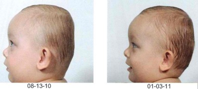
2. Brachycephaly

Brachycephaly is a flattening of the whole back of the skull. This result in a very wide head shape that appears wider compared to its height. The forehead may also be prominent to compensate with the flattening of the back of the head.

Brachycephaly Image

Brachycephaly Image

3. Scaphocephaly or Dolichoceophaly

These conditions occur when the head is longer in relation to its height. These usually result from lying to one side for prolonged periods as seen in preterm babies in the neonatal intensive care unit. Side lying position allows for easy access of the neonate for feeding or monitoring purposes.

Dolichoceophaly Image

Dolichoceophaly Image

Plagiocephaly Causes

Plagiocephaly results mainly from positional factors. Plagiocephaly develops as a result of resistance or pressure on the soft skull. This can happen inside the uterus or may develop as the child grows. The most common causes of plagiocephaly include:

Restrictive intrauterine environment

Pressure inside the uterus resulting from other fetal structures such as the placenta causes the head of the fetus to flatten even when the baby is still inside the womb. Plagiocephaly becomes apparent at birth and resolves within two months.

Letting the baby lie in a certain position for prolonged periods

When babies lie on their backs all the time, it causes flattening of the occipital area because of constant pressure on the area. There has been an increase in flat head syndrome as a result of recommendations to keep babies lie on their back to prevent sudden infant death syndrome. Side lying for prolonged periods also makes the baby’s head flat on a particular side. This can also be seen in babies who are always on car seats, bouncy seats and carriers.

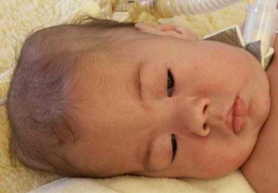
Torticollis

Torticollis or rye neck is the twisting of the neck which tilts the head to one side. This prevents the baby to turn their head because of neck pain. As a result, the baby assumes a certain position to prevent pain. Torticollis should be managed by physiotherapist because it causes considerable pain and may impair the breathing of the baby.

Torticollis Image

Torticollis as a cause of Plagiocephaly

Too little “cuddle time”

Letting the baby lie most of the time increases the risk for flat head. Caregivers should ensure to cuddle their babies once in a while to prevent plagiocephaly and improve bonding with the child.

Plagiocephaly Treatment

Plagiocephaly often do not need treatment because it can be resolved as the baby grows. However, treatments are also useful to improve the shape of the baby’s head and reduce anxiety of parents. Management for plagiocephaly focuses on preventing pressure on a particular part of the baby’s head. More serious cases may need corrective devices. These managements include:

Repositioning or Counter Positioning

Reposition the baby’s head during sleeping to prevent pressure at the back or at the sides. Avoid prolonged use of car seats and carriers, instead, cuddle the baby. Watch out for signs of torticollis such as crying when the baby’s head is moved to prevent further injury and pain.

Stimulate the baby to look at the opposite side of the flattening by using stimulating toys such as a big red balloon. Repositioning the baby’s head from time to time allows pressure on all areas of the skull and prevents flattening.

Cranial Remolding Orthosis or “baby helmet”

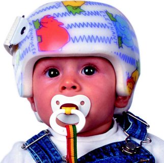
This device helps correct the abnormal head shape of babies by allowing the head to be contoured appropriately. The device is often used when repositioning does not improve the condition. It is also used in cases of positional plagiocephaly, scaphocephaly and brachycephaly. It is often prescribed for babies three to 18 months of age wherein the skull is still soft and can be remolded.

  • The cranial remolding orthosis provides pressure on the side that is more prominent and leaves a space on the other side to allow the head to be shaped normally.
  • Cranial remolding orthotics is light weight and made from a thin, hard shell lined with foam. It is worn 23 hours a day for maximum results. It can be taken off when the baby bathes. It is also readjusted from time to time by health care providers.
  • The use of cranial remolding orthosis is painless and takes three to four months to use the device. It has been effective in treating flat head syndrome over the years.
  • Parents should be instructed that this device is only temporary and has excellent outcomes.

Baby Helmet Image

Cranial Remolding Orthosis to treat Plagiocephaly

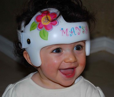
Custom Head Bands

Head bands provide a minimal amount of pressure on the baby’s head that needs reshaping. The band is usually provided and adjusted by pediatricians from time to time. It is more comfortable than the baby helmets, but is also prescribed for milder cases of plagiocephaly. Certain commercial head bands are also available for baby’s use. Regular head bands should not be used because it offers no relief from flattening of the head.

Presence of craniosynostosis is more severe and requires surgical management and advanced orthotics to correct the deformity. Fortunately, plagiocephaly does not require such.

Custom Head Bands Image

Custom Head Bands to treat Plagiocephaly

Plagiocephaly Prevention

How are we going to prevent plagiocephaly? Preventing the occurrence of “flat head syndrome” entails preventing too much pressure on a certain part of the head. These measures include:

  • Vary the baby’s position during sleeping time from flat, right side lying and left side lying position.
  • Try to change the position of the baby crib to prevent the baby from looking at certain things that keep their head to one position most of the time.
  • Reposition the baby in the crib such as changing the direction where his/her feet point to. This will also prevent the baby from positioning the head on certain areas because of constantly looking at a particular object in the room.
  • Place the baby on their tummy during playtime to prevent pressure on their soft head. This can be done four times a day.
  • Carry your baby from time to time to enhance bonding and prevent plagiocephaly.
  • Be sure not to put the bay in prone position or on his/her tummy during sleeping. Tummy time prevents plagiocephaly, but positioning in this way during sleeping may lead to suffocation and sudden infant death syndrome.
  • Avoid letting the baby sleep for prolonged periods in car seats and carriers.

Prognosis

Early studies have indicated that plagiocephaly results in learning difficulties and neurosensory problems. However, these studies have not been validated and experts now suggest that plagicephaly does not result to such. In fact, plagiocephaly do not lead to serious health problems.

The head deformity may be untreated because it usually resolves as the baby grows. Treatments discussed earlier are effective in managing plagiocephaly. Plagiocephaly does not need invasive surgical procedures to correct the deformity of the head.

Prognosis is excellent and most of the cases were naturally resolved before the baby’s first birthday.

Complications

Plagiocephaly does not result in complications aside from the lasting effect on the baby’s head. Plagiocephaly does not result in neurologic disorders or abnormalities in the baby’s brain development as well as sensory development. Facial asymmetry that results from plagiocephaly also resolves as the child grows.

No responses yet

IgA Nephropathy – Symptoms, Diet, Treatment, Prognosis, Causes

May 15 2012 Published by under Diseases & Conditions

What is IgA Nephropathy?

IgA nephropathy is a common disease that is a form of glomerulonephritis which is known as inflammation of glomeruli of kidney.

This is a condition where the IgA antibodies that fight against bacteria and virus will build up in kidney that will result in inability of kidney to filter waste, water and electrolytes that is an excess in the blood.

It’s also called by medical professionals as Berger’s Disease that if left untreated will eventually lead in having blood in urine, high blood pressure and swollen extremities.

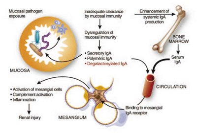
IgA Nephropathy Image

Process of IgA Nephropathy and its affectation

Signs and Symptoms of IgA Nephropathy or bergers kidney disease:

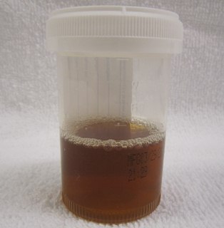
  • Blood in the urine that is normally tea colored

tea colored urine image

Tea colored urine as a symptom of IgA Nephropathy

  • The patient will also experience Upper Respiratory Infection
  • There is a visible swelling of the hands and the feet
  • The patient also experienced high blood pressure
  • Low-grade fever
  • Protein form like bubble in the urine
  • Nephrotic Syndrome that is caused by excessive loss of protein in the blood

Nephrotic Syndrome Image

Nephrotic Syndrome caused by excessive loss of protein

  • Extreme fatigue due to anemia
  • Flank pain on the sides due to inflammation caused by IgA Nephropathy

Causes of IgA Nephropathy

The main cause of disease or process on how it was deposited in glomeruli of kindney is still unknown. The causes that are mentioned can be associated in the formation of IgA Nephropathy.

  • Genes
  • Cirrhosis is a disease that will cause formation of scar tissue in the kidney that might affect the IgA to be deposited in the kidney
  • Celiac Disease is a condition where the person who has this cannot tolerate gluten that is common on breads that can cause for having blockage that will result in the deposition of IgA in the kidney.
  • Bacterial and Viral infections

Diagnosis of IgA Nephropathy

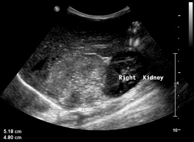
  • Ultrasound of the kidney can help the medical professionals to view and determine if there are present abnormalities in the kidney.ultrasound of kidney image

Renal Ultrasound to detect kidney abnormalities

  • Cytoscopy is used by medical professionals to examine the bladder and the urethra to check where the bleeding has started or other abnormalities that cause the bleeding.
  • Urine Sample to help the medical professionals to identify presence of blood and protein in the urine.
  • Blood Test for the medical professionals to identify the levels of waste products in the blood like if creatine level and the blood urea nitrogen level are high it is indicated that there is a problemin the kidney function.
  • If there is positive damage in the kidney, biopsy is recommended by the medical professionals. Biopsy can determine the damage present in the kidney and it can show presence of IgA deposits in the kidney. Biopsy is a medical type of test done by the medical professionals where they will get sample tissue used for examination. The sample that is removed is checked through a microscope and it helps to determine any present diseases.

Treatment and Diet for IgA Nephropathy

The goal of treatment for patients with IgA Nephropathy is to relieve the visible signs and symptoms and prevent renal failure. These are the treatments used for patients with IgA Nephropathy:

  • Medications that help to control blood pressure. These medications can help to delay and prevent kidney damage. Medications like Angiotensin-Converting Enzyme and Angiotensin-Receptor Blockers are the common medications that is ordered by medical professionals to take by the patients with IgA Nephropathy to decrease blood pressure.
  • Medications that will help patients to decrease cholesterol levels. Lowering cholesterol levels of the patients with IgA nephropathy can help the patients to slow the development of kidney damage.
  • The medical professionals also give immunosuppressant drugs like corticosteroid that will help to protect the kidney of the patient.
  • If kidney failure is present to the patient, the medical professionals will recommend Dialysis treatment or if possible Kidney transplant is also recommended. Dialysis is a procedure that does what the normal kidney do like removal of fluid and waste that people with kidney problem cannot do due to the damage of their kidney.

Diet for People with IgA Nephropathy

  • Low protein diet because protein rich foods will make the person glomeruli to function without rest thus making the kidney pressure higher.
  • Medical professionals recommend the patient with IgA Nephropathy to be a vegetarian. To prevent or slow the progression of kidney damage.
  • Diet that has low salts as well as controlling fluid intake of the person is also part of the treatment to reduce the swelling that the person is experiencing.
  • These foods should be avoided by the patient:
  • Stimulating foods such as seafood, beef and spicy foods.
  • Foods that stimulate the internal heat of the body like chocolate and hot pepper.
  • Food that is high in potassium like banana, potato and tomato.
  • Patients that has a high uric acid should avoid foods like fish, shrimp, and crab.

Prognosis of People with IgA Nephropathy

The prognosis of this disease depends on the health status of the patient. Usually the progression of this disease is slower except if the person who has an IgA Nephropathy has a high blood pressure, large amount of protein that is present in the urine and has an increase in Blood Urea Nitrogen Exam that indicated problem in the kidney function can make the progression faster and became worst because once the patient has a kidney failure it is irreversible.

Complications of IgA Nephropathy

  • High Blood Pressure :patients with high blood pressure will increase their risk in damaging the kidneys that will result:
  • Renal Failure : Kidney that is present in our body has a major contribution in keeping the body healthy. They are responsible in filtering blood, removing waste products, and they are also responsible in controlling blood pressure in the body. When there is damage in the kidney, the filtering ability of it will looses that will result in the buildup of waste in the blood thus resulting for the patient to undergo dialysis that will help in cleaning and removing waste in the blood.
  • Nephrotic Syndrome : A disease that is caused due to damage in the glomeruli that is responsible in controlling protein. In this disease the problem is the excessive release of protein substance in the urine that can result for the person to swell and contribute in the kidney damage.
  • End-stage Renal Disease : A disease that the kidney has the complete inability to function. Usually the medical professional recommend the patient to undergo kidney transplant and these patients will undergo continuous dialysis to remove the build-up of waste products in the body.

No responses yet

Next »

© 2017 eHealthWall.com. All Rights Reserved. Privacy Policy
This website is for informational purposes only and Is not a substitute for medical advice, diagnosis or treatment.