Archive for May, 2016

Osteogenesis Imperfecta

May 31 2016 Published by under Diseases & Conditions

What is Osteogenesis Imperfecta?

Osteogenesis imperfect (OI) is a bone disorder involving genetic predisposition. It is also called as Lobstein syndrome or brittle bone disease. Individuals with osteogenesis imperfect lacks Type-1 collagen, which leads to defects in the connective tissue or may also lead to inability to make connective tissues leading to brittle bones.

osteogenesis imperfecta

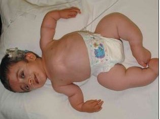
Type III Ostegenesis Imperfecta

Image source: pediatricsconsultant360.com

Types of Osteogenesis Imperfecta

Osteogenesis imperfect include eight types. There are varying severities as well as the gene involved in these types of the disease. These are:

Type I

Type I OI is the most prevalent type and it is considered the mildest form of OI. The structure of the collagen is usually normal, but there is insufficient number of the collagen leading to weakening of the bones.

Type II

Type II OI is considered the most severe type of OI. This is due to the defects in the collagen formation. Patients with this type have severe brittleness of the bones that may result to fractures even before birth. This type usually does not support life and fetus may not survive.

Type III

Type III OI involves adequate collagen, but they are improperly formed causing bone deformities and other complications.

Type IV

Type IV OI is considered moderately severe. Just like Type III, the collagen is of sufficient quantities, but has poor quality.

Type V

Type V OI has similar features than Type IV. Type V has characteristic mesh-like appearance of the bones under histological examinations.

Type VI – Type VI is also similar to Type IV with a characteristic of fish-scale bone.

Type VII  – This type is recessive in nature and was recently discovered.

Type VIII – Type VIII is similar to other types similar to LEPRE 1 gene.

Osteogenesis Imperfecta Symptoms

The severity of symptoms often differs according to type. Common symptoms include:

  • Weak bones
  • The classical symptom of osteogenesis imperfect is weak bones due to defects in the collagen matrix that helps strengthen the bones. Patients usually suffer from fractures even in the absence of high-impact tension.
  • Short stature
  • The growth of the long bones is also impaired because of collagen defects.
  • Breathing difficulties
  • The bones in the rib cage may be weak leading to inability to expand during inspiration.
  • Triangular-shaped face
  • The cranium may also be affected leading to problems in the formation of the facial bones.
  • Hearing loss
  • The small bones in the ears responsible for vibrating and transmitting sound waves is also weakened and may be fractured leading to permanent hearing loss.
  • Bone deformities
  • The weak bones is unable to support the normal structure and position of the bones and may also not be able to efficiently bear the weight of the patient leading to deformities such as scoliosis and bow-leg.
  • Brittle teeth
  • The teeth are made up of strong collagen matrix in order to become strong. As a result of OI, the teeth may become weak and prone to cavities.

Osteogenesis Imperfecta Pathophysiology, Genetics, Causes

Osteogenesis imperfect results from autosomal dominant inheritance of gene defects of connective tissue formation. However, more recent studies have also found some cases of autosomal recessive in nature. OI can be inherited from parents, but some cases may also be a result of sporadic or individual mutation without the factor of inheritance.

The defect in the Type-1 collagen gene results in the substitution of the amino acids to a bulkier amino acid in the collagen helix. Because of the larger amino acids, there is hindrance that creates a bulge in the collagen creating a defective collagen structure. The presence of improper collagen makes the structure of the bones brittle.

Genetic predisposition is very strong; however, some patients may have developed the gene mutation themselves and may stop functioning adequately.

Osteogenesis Imperfecta Diagnosis

The diagnosis of OI starts with a thorough physical examination including the teeth and the eyes. Presence of family history of osteogenesis imperfect may also be determined. Other tests essential for diagnosing OI include:

X-rays

X-rays provide a clear image of the bone structure and other malformations. Patients usually exhibit one or more stress fractures on the skeletal system.

Genetic Testing

Genetic testing may be performed to identify the specific genetic mutations.

Ultrasound

Ultrasound may be also performed when the mother is pregnant to determine OI in utero. Severe cases of OI may be detected from the fetus using ultrasound.

deformed long bone x ray


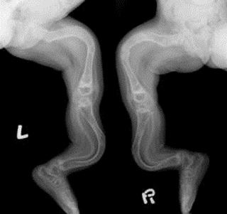
X – ray imaging showing long bone deformity

Image source: physio-pedia.com

Treatment

There is no cure for osteogenesis imperfect because of the genetic mutation. Treatments involved are only supportive to enhance the bone strength, maintain mobility and prevent fractures. These include:

Physiotherapy

Physiotherapy is used to improve the mobility and strengthen the muscles thereby enhancing the shock absorbing property of the muscles to prevent bone injuries.

Exercise

Regular weight –bearing exercises help in increasing the bone density of patients.

Proper diet

Diet rich in calcium and vitamin D is essential in improving the bone density and prevent further osteoporosis. Vitamin D and calcium supplements may also be given.

Mobility aides

Mobility aides are used such as wheelchairs, crutches and grabbing arms to improve autonomy and mobility of patients.

Biphosphonate therapy

Biphosphonates in the form of oral alendronate or intravenous pamidronate and zoledronic acid are also increasingly used for OI to increase the bone mass. Side-effects of these medications include nausea, bone pain and dizziness. However, therapy with pamindronate can also lead to weakness of bones so regular bone density determination is required.

Surgery

Surgery serves as a last resort for the treatment of OI. Metal rods can be inserted in the long bones to increase strength and serves as support. Metal rods also increase in length as the child grows to support the growing bones.

In cases of severe scoliosis, spinal fusion may be done to correct the spinal deformity. Spinal fusion involves the realignment and fusing of the spinal bones.

Prognosis

Prognosis of OI depends on the type and severity of the symptoms. Patients with Type I have a better prognosis. On the other hand, those that have Type II OI may not survive at birth. The most common cause of death is respiratory failure and accidental trauma. Those who are treated often live normal lives.

Life expectancy

The life expectancy of patients with OI is indefinite; however, statistics shows that more than half of cases die before the age of ten. Those who reach more than this age usually have better outlook.

No responses yet

Mastoiditis

May 23 2016 Published by under Diseases & Conditions

What is Mastoiditis?

Mastoiditis is a termed used to describe an infection that is located at the person’s mastoid process, which is found on the temporal bone located on the skull just behind the person’s ear. It is often due to otitis media or infection of the person’s middle ear. The infection associated with Mastoiditis is bacterial in nature.

The infection occurs within the mastoid process but some cases have an ear infection which spreads throughout the mucus membrane that surrounds the mastoid itself until it reaches to the bones, which is far graver when reached within this case. It is used to be a leading etiological factor of the death in most children. It often starts with a small infection which progresses into a life-threatening problems or complications.

Mastoiditis Symptoms

Patients with Mastoiditis will manifest the following symptoms associated with the disease condition such as:

  • Headache
  • Pain in the ear or otherwise known as otalgia
  • Irritability
  • Tenderness of the ear specially in the mastoid region
  • Drainage from the person’s ear which may be brownish in color
  • Swelling of the ear
  • Erythematous
  • Infection of the ear
  • Fever
  • Vomiting
  • Chills
  • Anorexia
  • Lethargy
  • Drooping and bulging of the person’s ear
  • Diarrhea or loose bowels

These are the common symptoms associate with this disease condition. The non specific symptoms such as diarrhea, anorexia, irritability or lethargy may be shown in infants.

Mastoiditis Terminology

Acute Mastoiditis

It is a common disease category of Mastoiditis that occurs most often in children. When one neglects it, it may progress to Chronic Mastoiditis. It is a common occurrence before but today with the advancement of medical treatment, it occurs very rare and may be seldom seen. When one has this, the person has a history of infection of the ears. The cardinal sign for this condition is pain. Even after acute form of otitis is treated, the pain will typically subside. However, if the pain worsens, then the person will positively have infection of the mastoid. The inflammatory process will then be manifested which is the dolor, calor, pallor, rubor, function laesa and tumor.

Chronic Mastoiditis

When person will acquire chronic form of Mastoiditis, it usually means that they may or may not have chronic for of otitis media. They have the following symptoms: low grade fever, tenderness and chronic otalgia. This kind of classification of Mastoiditis occurs when the person does not respond well to the treatment or perhaps it recurs often.

Bilateral Mastoiditis

It is a medical term used to describe infection on the mastoid portion which affects both your ears. It may either be in a form of acute or chronic kind. It is due to an untreated kind of infection of the middle ear portion.

Coalescent Mastoiditis

It is a medical term given to persons who have acute form of otomastoiditis. It happens when mucoperiosteal kind of disease that involves the bone. It is the rare kind of infection of the mastoid bony portion or process. When the disease doesn’t respond well to treatment, it may have the possibility to spread to the person’s brain which will then result to disability or worse, to death.

Left Mastoiditis

When a person has this diagnosis, it means to say that the person has an infection of the mastoid portion which is located behind the person’s ear on the left portion. The right ear is unaffected.

Mastoiditis Causes

As mentioned earlier, persons who are afflicted with Mastoiditis have bacterial form of infection. Patients who have the disease condition often were exposed or have a medical history of ear infection, particularly in the middle ear, which may often recur. It affects children. Aside from that, those persons who have weaker immune system may be afflicted with the disease.

Mastoiditis Diagnosis

The physician will conduct examinations to be able to diagnose persons who are suggested to have Mastoiditis. The examination that the patient undergoes includes the following:

  • Physical examination
  • Medical history examination
  • X-ray examination which may or may not be useful but some physician would use it
  • Blood test examination
  • Ear Gram staining culture examination
  • Biopsy
  • MRI scan
  • CT scan
  • Technetium 99 bone scan examination
  • Exploratory Surgery for diagnosis

It is suggested that when either of the symptoms above mentioned will be present, there is a need for you to consult a trusted physician as early as possible before the condition becomes worst and treatment will not be any more effective.

Ct scan showing bon e infection

CT scan showing bone infection located at left mastoid process

Mastoiditis Treatment

Depending on the extent of the disease condition, the person with Mastoiditis is at the most treated by either:

  • Antibiotics to treat the bacterial infection and which may be done intravenously which is the fastest route to use. Oral antibiotics will be given after the intravenous antibiotic is given.
  • Antipyretics to treat fever episodes that are brought about by the inflammatory process or in the presence of fever.
  • Myringotomy which may prevent ear infection in the future and Mastoiditis that appears subsequently. It is done to drain the ear fluid found in the middle ear portion.
  • Mastoidectomy which is a surgical procedure done especially when the treatment mentioned above is unsuccessful. Here, the mastoid portion is removed to prevent the spreading of the infection to the nearby areas.

Mastoiditis Complications

When undergoing a serious surgical procedure, you must outweigh advantage and disadvantage to consider, for it can result to serious complications such as

  • Blood clot
  • Brain abscess
  • Meningitis
  • Hearing loss
  • Bell’s palsy
  • Osteomyelitis
  • Otitis media
  • Cellulitis
  • The mastoid bone is destroyed
  • Vertigo or dizziness
  • Facial paralysis

Hence, do not wait for this to occur. Although most of the complications are of rare occurrence, there is a need to seek treatment promptly.

One thing is for sure, is that the complications which are associated with this disease condition are rare. However, the complications will occur if the person does not seek immediate medical care and if the person does not respond well to the treatment provided.

No responses yet

Best Recipes for Weight Loss: What to Look in a Healthy Recipe?

May 15 2016 Published by under General Health

Having to lose weight is one of the greatest problems of most people, which until now, has not provided a quick ad fats result of losing weight. This is due to the fact that food is a basic necessity and avoiding it is just too difficult for some. Also, the foods we have now seem to have caused obesity more rater the foods we had decades ago. Because of the advent of technology and the rise of preservative and a lot of additives to our food to make it taste better, chances are the food we eat becomes less and less healthy. In order to address this and maximize weight loss, it is best to choose the recipes we are going to cook.

recipes for weight loss

How to Choose the Best recipes for Weight Loss

The following are the tips in choosing the best recipes to make our food taste better and help us lose weight.

Check the ingredients rather than looking at the pictures

Most of us look into recipe photos to choose what we are going to cook. Chances are, the most appetizing foods are the unhealthiest ones. Next time you pick recipes, better read the ingredients and chose healthy choices. Also, check the cooking methods. Choose the recipes that require only boiling, blanching or broiling rather than frying because frying adds oil, which is high in fats.

Choose all natural ingredients

When deciding to cook, choose all natural ingredients. Instead of using canned or preserved veggies and meats, use the fresh and natural ones because they contain less fat, sodium and other additives. If you will cook noodles, try to make your own noodle pasta rather than buying the commercially prepared ones. In this way, you are assured that what you are eating strictly comes from nature.

Avoid those recipes with most meats in it

As meat is essential to get protein, pure meat diet or recipe is also not good. Meat contains more cholesterol and fat that make you unhealthy, plus it is more difficult to eliminate. Instead of choosing these recipes, choose those who contain more fruits and vegetables.

Avoid all carb recipes

Carbohydrates are the primary source of energy, but taking too much of carbs will only make you fat especially when you are not engaging in an active lifestyle. Instead of all carb recipes, try to balance it by adding proteins, fruits and vegetables.

Reinvent the recipes

Recipe books do not say that you have to stick to the instructions and ingredients. If you want something, but fear that this can only make you fat, then you can reinvent the dish. For instance, instead of preparing white pasta spaghetti, why not use whole wheat pasta, which is healthier and less fattening than regular pasta. Also, instead of frying your favorite fish, you can just steam it the way you want.

Check your calorie intake

Before starting to cook any dish for the day, you have to set a goal on the total calories you need to have. In this way, you can spread your calorie intake throughout the day. Also, choose recipes that may fall into the calorie intake required for a specific meal. This may sound difficult and taxing because you need to compute everything. However, you can use the weight loss Apps online, which already provide you a meal plan for a specific day depending on your caloric need. You just have to choose the best recipes it provides using the above tips.

When you are able to follow these simple steps, you already lose weight in no time.

No responses yet

Esotropia – Pictures, Definition, Surgery, Treatment

May 07 2016 Published by under Diseases & Conditions

What is Esotropia?

Esotropia, a form of strabismus, which occurs when there is a misalignment of the eyes which is distinguished as a convergence of the visual axes which is done excessively leading to an appearance of “cross eye”. What happens is that the lateral muscles of the rectus are paralyzed which resulted to one eye being abnormally deviated inwardly, towards the person’s nose, which is observed during an attempted gaze.

There is actually visual signal suppression from the eye to the brain which results to having the decline depth of perception. While the other eye functions normally. This is a common ocular disorder found in infants.

Esotropia Image

Misalignment of the eyes or Esotropia

Symptoms of Esotropia

Persons diagnosed as having Esotropia are unable to use both eyes together especially when gazing at a particular object. The popular symptoms manifested by a person having this ocular disease condition are as follows:

  • Double vision or strabismus
  • Decreased vision
  • Misalignment of the eyes

Aside from this, you might observed that persons with this ocular condition, are found to squint often during bright sunlight or are found to tilt their heads towards one direction just to be able to use their eyes together. They often will rub their eyes in a repeated manner. Children, who comprise the most numerous number of reported esotropia, will at times not be able to tell you that they are experiencing the symptoms mentioned. However, you might observe that they close one eye, usually the eye that is impaired, to be able to compensate for the visual problem. You might also notice a faulty perception to the objects that are presented to them.

The brain of a child, having double vision or strabismus, ignores the image of the misaligned eye and will only constantly use the other functional eye. This is called as the lazy eye or amblyopia. They often experience such condition associated with this kind of ocular problem. However, with adults, lazy eye is easier to pinpoint since the brain is trained to accept the images coming from both eyes and it can’t ignore the image from the impaired eye.

Causes of Esotropia

This ocular condition may be due to the fact of the following the common reasons:

Refractive Errors

There is an occurrence of refractive errors because the focus of the light by the eye is incorrect.

Insufficiency in Divergence

There is an insufficient divergence since the person doesn’t see the distant objects clearly.

Excess Convergence

There is excess in convergence or what is termed as strabismus, is usually seen when the eyes fail to look at the same direction.

Other causative factor that may lead to having esotropia includes:

  • Damage to the sixth cranial nerve
  • Genetic disorders
  • Optic Nerve Hypoplasia
  • Deficient in Glycerol Kinase
  • Bilateral Frontoparietal Polymicrogyria
  • Carbohydrate deficiency in Glycoprotein Syndrome type 1a
  • Tumor

Diagnosis of Esotropia

The physician usually does the diagnosis of this ocular condition. What the physician usually does is that he or she first identifies this disease as a pathological. The first six months, is when the infantile esotropia develops. As stated above, esotropia is usually found in infants. However, the disease may be transient, going on and off, which will delay diagnosing this ocular problem.

Second, the physician will measure the eyes through the alternate prism test. It will measure the angular deviation of the eyes. An angle of 20 prism diopters means that the patient is positive with infantile esotropia. When it falls between 20 to 40 prism diopters, there is still a chance to resolve the ocular problem. However, when the angle falls to 40 prism diopters and above, it is expected that it will rarely resolve by itself.

Prism Image

Alternate prism test to measure the angular deviation of the eyes

Third, the physician will check for amblyopia or for crossing of the eyes. After that, he or she will check for additional signs of ocular motor problems such as incomitance, impaired binocularity, and scotomas. Lastly, the physician will look for other additional underlying etiology of esotropia.

It is important to note that the physician will do tests to be able to know the strength of the muscles located in the extraocular area. Such test includes:

  • Visual acuity
  • Standard ophthalmic exam
  • Retinal Exam
  • Neurological Examination

Esotropia vs. Esophoria

The main difference between Esophoria and Esotropia is that in Esophoria, the eye is deviated inwardly with is associated with an imbalance in the extra-ocular muscles. It is a convergent strabismus or a squint. It is often constant and is reported to be present before the school age, usually in the ages 2 and 3 years old. Meanwhile, Esotropia, there is a possibility of fusion and diplopia or having double vision is uncommon with this disease.

Treatments of Esotropia

There are actually many options in treating Esotropia. This treatment can be either:

  • Optical Correction – Most often, this optical problem can be primary treated with the use of corrective glasses. The ophthalmologist, a physician specialized in vision and eye care, will prescribe the glasses that is designed to treat the impaired eye.
  • Optical correction for esotropia

Optical correction for esotropia

  • Added Lens Power – Some practitioners would suggest overplussing the child. Overplussing the child is done through using an additional plus power added to the singular vision lens.
  • Prisms – As mentioned earlier, this test will measure the angular deviation of the eyes.
  • Active Vision Therapy – It is done through the use of penlights and mirrors, biofeedback, tracing pictures, puzzle completion, and video games. Electronic optical instruments may be used to enhance the therapy.
  • Pharmacological Agents – Some Physicians will prescribe medications to treat Esotropia; such of this treatment includes miotic medications.
  • Chemodenervation – Chemodenervation treatment is usually done by injecting Botox or Botulinum Toxin. It is injected in the extraocular muscles. It will help in relieving the strabismus found in Esotropia

Chemodenervation treatment

Chemodenervation Treatment

  • Surgery – Lastly, if all the other treatments mentioned didn’t succeed, surgery is considered the last most effective option. The surgeon will correct the underlying disorder by correcting it through surgical operation.

No responses yet

© 2017 eHealthWall.com. All Rights Reserved. Privacy Policy
This website is for informational purposes only and Is not a substitute for medical advice, diagnosis or treatment.