Archive for June, 2016

Erythema Multiforme

Jun 30 2016 Published by under Diseases & Conditions

What is Erythema Multiforme?

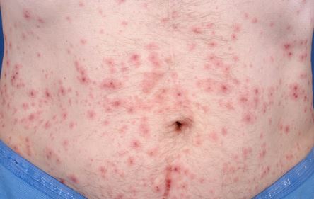
A skin condition proposed to be a result of hypersensitivity reaction from drugs or infectious agents. erythema multiforme is characterized by red to pinkish papules, macules and the hallmark “target lesions” that are distributed throughout the limbs and sometimes the mucus membranes.

erythema multiforme picture

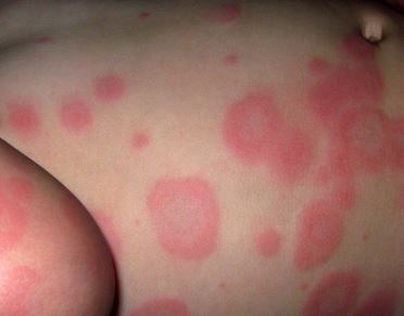
Image 1 –  Erythema multoforme on body

When Does Erythema Multiforme Happen?

The red (erythema) rashes usually emerge after exposure to a drug or an infectious agent, for this erythema multiforme is considered as an allergic reaction to a particular agent.

Erythema Multiforme Presentation

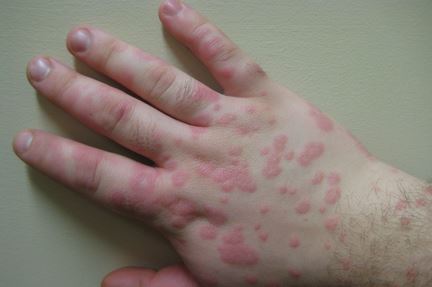
The emergence of the rash would vary from a self-limited rash to a life-threatening, severe one. Rash can present itself as small pink macules or papules, and as the condition progresses, the rash becomes large and widely distributed. Erythema multiforme rashes are usually located on the extremities, and rarely may involve the mucous membranes.

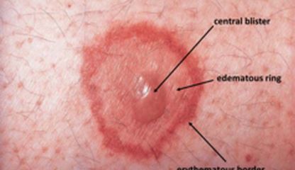
The hallmark sign of multiform erythema rash is its target like appearance. Here the lesions appear pale on the center, and pink-red around the circumference. It can also be itchy (as in mild cases) and can cause abrasions and scratches in severe cases when the itching escalates.

Erythema Multiforme Symptoms and Signs

  1. Itching or burning sensation on the site of the rash
  2. Numerous red-pink maculo-papular rash that begins on the extremities then may proceed to the torso
  3. Sharply demarcated pink to red rash
  4. Presence of a target like lesion (also called “iris” lesion)
  5. Presence of plaques
  6. Plaques or papules that have a darker, brown, purpuric, center (happens at the late stages of the condition)
  7. Presence of blisters and crusting on the center of the lesion may also be noted.
  8. Presence of mucosal lesions especially in the mouth

Other Signs and Symptoms may include:

  1. Fever
  2. Muscle pain
  3. Body malaise or weakness
  4. Cough or sore throat
  5. Joint pain

Erythema Multiforme Causes

Since this condition is a hypersensitivity reaction, the following agents are seen to cause erythema multiforme:

  • Herpes Simplex Virus (HSV) – This is the most common agent seen to cause erythema multiforme, almost 50% of the cases are attributed to infection of the herpes simplex virus. HSV has the capability to express its DNA on skin cells, particularly keratinocytes. When their genes are expressed in these cells, our body’s defense system forms killer cells to destroy the viral genes within the skin cells since it is a foreign body. The destruction of the HSV gene also causes the keratinocytes to be destroyed, and this process sparks the inflammatory reaction we seen in erythema multiforme.
  • Mycoplasma pneumoniae – Another infectious agent seen to cause erythema multiforme is Mycoplasma pneumoniae. This is an agent that causes fungal infections especially in children. The toxins released by this fungal agent may be the cause of rash and lesion formation seen in erythema multiforme.
  • Medications such as:
  1. Barbiturates
  2. Penicillins
  3. Sulfa drugs or sulfonamides
  4. NSAIDs or non-steroidal anti-inflammatories
  5. Hydantoins
  6. Phenothiazines
  • Vaccines for:
  1. Hep B
  2. Small Pox
  3. Hepatitis C
  4. Varicella Zoster
  5. Cytomegalovirus
  6. Human immunodeficiency virus
  7. Dipthteria-tetanus
  • New medications are also related to erythema multiforme formation:
  1. Rofecoxib (Vioxx)
  2. Candesartan cilextil (Atacand)
  3. Ciprofloxacin (Cipro)
  4. Metformin (Glucophage)
  5. Bupropion (Wellbutrin)
  6. Adalimurab (Humira)

Erythema Multiforme Diagnosis

Clinical Assessment

Simple assessment of the patient for the presence of target lesions is enough to diagnose erythema multiforme. Further strengthening the diagnosis is the presence of an HSV infection

Skin Biopsy

This is done when the clinical manifestations are not clear. For patients with recurring erythema multiforme or for those with no history of HSV infection, skin biopsy may be done.

Laboratory Tests

Laboratory tests such as Immunoglobulin M and G, HSV-1 and 2 may also be done if the clinical presentation is unclear.

Erythema Multiforme Differential Diagnosis

Many conditions also resemble the clinical presentation of erythema multiforme. Differential diagnosis is important to rule out other forms of condition from erythema multiforme. Differential diagnoses for this condition include the following:

  • Urticarial and urticarial vacuities
  • Vasculitis
  • Viral exanthems such as measles
  • Polymorphic light eruption
  • Lupus erythematosus
  • Pityriasis rosea
  • Steven-Johnson’s syndrome
  • Drug eruption
  • Toxic Epidermal necrolysis

It is important for the physician to take note of the distinguishing characteristics of erythema multiforme. This condition is usually fixed and present for at least one week, and lesions have the characteristic “iris” appearance or the so called “target-like” lesions. Other lesions only emerge for less than 24 hours (urticaria), and do not resemble the target appearance of erythema multiforme.

Erythema Multiforme Treatment

Oral Antihistamines and Steroids

This relieves the symptoms of inflammation such as redness and itchiness. These medications work by suppressing the release of chemicals that cause the inflammatory reaction of redness and itching.

Side effects of antihistamines and steroids

Antihistamines can cause dizziness, and so it must not be taken with alcohol or sleeping pills. Short term steroid use can cause sudden weight gain, facial flushing and increase in appetite. If steroids are used for a long period of time, gastric acidity and immunosuppression are the common side effects.

Oral Acyclovir

Oral acyclovir is given to patients with ongoing HSV infection. Acyclovir is an antiviral drug that destroys the virus and prevents it from replicating. Acyclovir lessens the duration of the eruptions and so is the number of lesions

Side effects of Oral Acyclovir are he following:

  • Nausea
  • Abdominal pain
  • Vomiting
  • Dizziness
  • Confusion

Prednisone

Prednisone 40-80 mg/day is given for patients with multiple lesions. Its dosage lessened gradually as the lesions subside. Prednisone has an anti-inflammatory effect, suppressing the immune response that causes the inflammation we see in erythema multiforme

Side effects of prednisone

  • Water retention
  • Insomnia
  • Abdominal pain
  • Osteoporosis
  • Facial swelling

Valacyclovir and Famciclovir

This is given for patients with recurring condition and does not respond to Acyclovir treatment. A dose of Valcyclovir 500-1000 mg and Famciclovir 120-250 mg per day can be given. These are antivirals which halt the viruses’’ capability to strike damage to the cells.

Dapsone treatment

This is given for those with recurrent erythema multiforme who are not responsive to antiviral therapy. Dapsone 100-150 mg per day is given and is seen to partially and completely relieve erythema multiforme. Dapsone has anti-inflammatory and immunosuppressing action, this mechanism helps in suppressing erythema multiforme.

Side effects of Dapsone

  • Headache
  • Possible hemolysis (related to doses given)
  • Nausea
  • Peripheral neuritis

Azathioprine or Immuran

This is given if other treatments were not effective; Azathioprine has immunosuppressing effects, modulating the overactive immune response we see in erythema multiforme. This is a potent drug which suppresses the production of chemicals that spark the immune response.

Side effects of Azathioprine or Immuran

  • Susceptibility to other infections (because of bone marrow suppression)
  • Fatigue
  • Hair loss
  • Nausea
  • Can be a carcinogen if used as a long term treatment

Erythema Multiforme in Children

In children, erythema multiforme is commonly caused by an infection rather than exposure to medications. Primarily causing erythema multiforme in children is the Herpes simplex virus and Mycoplasma pneumoniae. Since this is an infection, erythema multiforme in children is most often coupled with other symptoms such as fever, body weakness, sore throat, muscle pain and joint pain.

Is Erythema Multiforme Contagious?

No, erythema multiforme itself is not a contagious disease. This is a condition that is caused by an allergic reaction to medications or an inflammatory reaction to viruses and fungi. Erythema multiforme is not indicative of a contagious disease and must not be mistaken as a sign of measles and other viral exanthems.

Erythema Multiforme Terminology

Erythema Multiforme Major

This is a severe life-threatening form of erythema multiforme; this type can be related to Steven-Johnson’s syndrome. A condition characterized by severe allergic reactions. Severe allergic reactions can cause anaphylactic shock, the event where a person’s airway is blocked and the blood pressure drops due to severe allergic reaction.

Erythema Multiform Minor

This is the less severe and most common form of erythema multiform. This is highly associated with a Herpes simplex infection. This form is only limited to cutaneous lesions, and is not systemic as compared to erythema multiform major.

Bullous Erythema Multiforme

This type of erythema multiform is characterized by bullous lesion formation. A bullous lesion is a type of a large lesion that contains fluid. In bullous erythema multiform, the rash may initially appear small and flat; as the condition progresses the lesions enlarge and resemble vesicles that contain fluids. This is called a bullous lesion.

Recurrent Erythema Multiforme

If the lesions are treated, but re-emerges again after sometime, the condition is called recurrent erythema multiforme. This usually happens as result of HSV infection which was unresponsive to the previous treatment. Suppression of the virus and prophylactic treatment of acyclovir is employed to prevent recurrent erythema multiforme, but in some cases higher dosage of antivirals and dermatological treatments are needed to prevent recurring episodes.

Erythema Multiforme Pictures

Erythema multiforme image

erythema multiforme pic

erythema multiforme on hands

No responses yet

Myelosuppression

Jun 25 2016 Published by under Diseases & Conditions

What is Myelosuppression?

Myelosuppression otherwise known as bone marrow suppression is a reduction in the capacity of the bone marrow to generate blood cells. Bone marrow suppression results in decreased numbers of circulating leukocytes (leukopenia), erythrocytes (erythrocytopenia), and platelets (thrombocytopenia). Decreased leukocyte numbers cause immunosuppression. Decreased erythrocytes and platelets cause hypoxia, fatigue, and increased bleeding tendency. Myelosuppression can be mild to severe (fatal) based on the number of cells produced.
Most chemotherapeutic agents suppress bone marrow function to some degree. The agents associated with severe bone marrow suppression include busulfan, cyclophosphamide, cytosine arabinoside, dactinomycin, doxorubicin, daunorubicn, etoposide, mitomycin C, nitrogen mustard, and triethylenethiophosphoramide. Immunosuppression, which places the client at extreme risk for infection, is the major dose-limiting side-effect of cancer chemotherapy. Suppression of immune function is the main life-threatening side-effect and presents the nurse with serious challenge of providing the client with understanding, environment, and support to withstand this potentially devastating Complication.

Myelosuppression Symptoms & Signs

The signs and symptoms of myelosuppression are largely related to the decrease in the levels of the blood components. Manifestations of myelosuppression include the following:

  1. Fever as a sign of infection
  2. The patient have bleeding tendencies and bruises (excessive bleeding) because of thrombocytopenia
  3. Increased risks of having infections because of leukopenia
  4. Symptoms of having an infection
  5. Fatigue because of anemia

Myelosuppression can result in diminished production of red blood cells, white blood cells and platelets. When the number of red blood cells decreases the patient may have anemia and easy fatigability. On the other hand, decrease production of white blood cells may cause leukopenia or neutropenia (decrease in the number of neutrophils), and when this happens the patient will be more prone to infection especially that neutrophils combat bacteria. The patient will be immunocompromised and his body will have a hard time combating infections. While when the platelets start to lessen it may cause thrombocytopenia and will result in bleeding tendencies and bruising.

Myelosuppression Causes & Risk factors

Causes and Risk factors for bone marrow suppression include:

Chemotherapy

Majority in all the cases the cause of myelosuppression is chemotherapy and other cancer treatments. This is because a lot of the medicines used in chemotherapy momentarily repress the bone marrow.

Therapeutic x-rays

These are also devastating when it reaches the bone marrow. The radiation combats the function of the bone marrow in producing RBC, WBCs and platelets when given in large proportions in increased frequency.

Cancer

The cancer cells themselves can also lead to myelosuppression. A number of cancers attack the bone marrow and squeeze out the cells usually residing there. However, there are others that can suppress the bone marrow without attacking them directly such as deficiencies in nutrition, which is very frequent in people who suffer from cancer, gradually slow production of the blood cells as well as viral infections and several non-chemotherapeutic medications.
The condition more often than not initiates more than a week following damage to the bone marrow. Nevertheless, the bone marrow usually goes back to normal throughout the next several weeks. Not as much of, increasing injury can be brought about by infrequently, permanent damage that causes irreversible myelosuppression. Extremely rigorous radiation, chemotherapy or other cancer treatments have the ability to demolish every one of the cells in the bone marrow.

Myelosuppression Diagnostic Tests

Laboratory tests employed for the diagnosis of myelosuppression include the following:

Blood Count Tests

Having the blood extracted is a common practice among cancer patients. Especially that myelosuppression is one of the side-effects of the chemotherapy, it is important for a routine blood test to de done in a regular way. A significant decrease in red blood cells, white blood cells and platelets should be reported to the doctor immediately. There are many lab tests to be done to find the red blood cell count, leukocyte and neutrophil count and platelet count and clotting function examinations. Anemia may initially be noticed using a Complete Blood Count (CBC). It is a usual test that calculates the number and relation of the proportion of every dissimilar kind of cells in your blood stream.

Bone Marrow Biopsy

When CBC is not that efficient the doctor may suggest bone marrow biopsy. This test also determines the presence of cancer cells in the bone marrow as a result of leukemia or other malignancies involving the bone marrow. This test is done by aspirating bone marrow cells on the ischium part of the pelvis. The aspiration is extremely painful so patient support is needed throughout the procedure.

Myelosuppression Treatment

Management of myelosuppresion includes treatments aimed to reduce the cause of myelosuppression as well as symptomatic management. A mild case of myelosuppression is not constantly managed.

Limiting Cancer Therapy

If the case is caused by chemotherapy or radiation therapy, the cancer treatments may be suggested stopping, lessening or postponing them. This is done to reduce the damage or to give the patient’s bone marrow a time to improve. The full dose of the cancer treatment should not be given.

Blood Transfusions

Meanwhile, the red blood cells and platelets can be simply replaced by blood transfusions (packed RBC’s or platelets). This will give a short-term solution to the problem and the bad news is the treatment may need to be done over and over because the transfused cells live only for a short period of time. There is also small risk of having a transfusion reaction and infection if the blood that is being transfused is contaminated by virus. In addition, sad to say, but transfusions of WBCs’ are not that effective and infrequently given by the doctors.

Growth Factor Therapy

Growth factor injections may be successful. Growth factors are defined as natural substances that can be found in the human body. This growth factor promotes blood cell production of the bone marrow. There are specific types of growth factor for certain blood cells, these are the examples:

  1. Erythropoietin injections
  2. granulocyte colony-stimulating factor
  3. granulocyte-macrophage colony-stimulating factor
  4. interleukin 11

The growth factor injections can lessen the need for blood transfusion and to make the life better for cancer patients. However, this has several side-effects if the kidneys are healthy and may not be successful if the body is producing enough erythropoietin. Other side-effects include pain in the bones, rashes, muscle soreness, nausea, and fever, retention of fluid, tachycardia, reddish eyes and dyspnea.

Bone Marrow Transplant

Bone marrow transplant is required if the bone marrow has permanent and severe damage. Bone marrows are taken from compatible donors.

Myelosuppression Complications

Complications of myelosuppression may include:

  1. Sepsis- Presence of severe infection as a result of very low white blood cells may eventually to systemic infection.
  2. Hemorrhage- The presence of thrombocytopenia or low platelet count results in poor platelet aggregation. This will eventually lead to profuse bleeding in all areas of the body including nose bleeding, hematochezia (bright red blood in the stool), severe bruising and the like.
  3. Severe anemia- Unmanaged bone marrow suppression often leads to severe reduction in RBCs. This condition results in excessive weakness, fatigability, difficulty of breathing and hypoxia.

Myelosuppresion Prevention

Prevention of bone marrow suppression depends on limiting or avoiding the cause such as:

  1. Exercising caution in administering chemotherapeutic agents.
  2. Avoiding exposure to high doses of radiation.
  3. Increasing the immune system by means of healthy diet, exercise and vitamin supplementation.

No responses yet

Jammed Finger

Jun 20 2016 Published by under Diseases & Conditions

What is Jammed Finger?

Jammed Finger is a condition wherein the finger suffers from an impact injury causing pain and swelling in the area. The condition tends to be very painful and requires further management. Jammed fingers relatively heal quickly when fractures are not incurred. Jamming of the fingers is more common, although affectation of the toes may also be experienced.

jammed finger image

When fractures are present, the healing time lengthens to one to two weeks and may require more extensive treatments. Jammed finger is a layman’s term for the axial blow on the proximal interphalangeal joint or at the tip of the fingers.

The occurrence of jammed fingers is more common in sports involving ball-handling such as volleyball and basketball. However, jammed fingers can be effectively prevented through training and special equipment in the fingers. Any injury in the proximal interphalangeal joint can lead to permanent disability when left undiagnosed and untreated.

The proximal interphalangeal joint is an axis joint that can perform range of motion from 0 to 120 degrees. The lateral stability of the joint is supported by the thick collateral ligaments. The injury in jammed fingers involves some degree of hyperextension in the area with longitudinal forces or compression on the tip of the fingers.

The volar plate forms the base of the joint. The joint also have extensor and flexor tendons that allow for various range of motion of the joint. The most common reason for jammed fingers is the axial blow or longitudinal compression on the joint with additional hyperextension of the extensor tendons that may result in dislocation of the volar plate. The injury may also affect one or more fingers, but the middle finger is most commonly affected.

Symptoms of Jammed Finger

Jammed fingers lead to various symptoms such as:

  • Tenderness on the proximal interphalangeal joint
  • Swelling or edema on the top of the finger or fingers
  • Limitation on the range of motion of the proximal interphalangeal joint.
  • Loss of active extension of the finger
  • Significant pain on the area
  • Numbness or tingling sensations in the finger as a sign of neurovascular compromise

Causes of Jammed Finger

Jammed finger can be caused by any activities that may involve longitudinal compression on the interphalangeal joint. These activities involve the use of the hand to handle things such as:

Sports

Basketball, volleyball, baseball, softball, and other sports may cause accidental longitudinal compression of the proximal interphalangeal joint during athletic activities. These causes are usually not preventable because injuries may happen during any sports events.

Falls

Accidental falls may also lead to jammed finger in cases that the fingers meet the ground first in a longitudinal manner, hyperextending and breaking the extensor ligaments in the fingers.

Diagnosis of Jammed Finger

Diagnosing jammed finger involves radiographic studies as well as physical examination to evaluate the condition of the patient and differentiate it from fracture of the fingers. These diagnostic tests include:

X-ray imaging

X-rays are very essential in diagnosing the condition and differentiating it from finger dislocations and fracture. X-ray images will reveal the exact damage on the proximal interphalangeal joint such as dorsal dislocations, volar plate sprain, collateral ligament injury and Boutonniere deformity. Radiographic studies involve the taking of lateral, anteroposterior ad oblique radiographic images of the digit. Radiographic studies are able to identify subluxation, dislocations and fractures.

Physical examination

Physical examination is usually done after radiographic studies to prevent further injury in cases of fracture or other conditions. Physical examination involves assessing for edema, swelling and tenderness in the area. The neurovascular status is also assessed by checking the skin temperature and presence of paresthesias or tingling sensation in the area. Finally, the range of motion of the joint is assessed, which may reveal loss of active extension of the joint.

Metacarpal block

This involves the injection of local anesthesia in the hands and fingers to reduce pain for more accurate diagnosis because pain may sometimes impede further assessment.

Treatment of Jammed Finger

How to fix a jammed Finger is a question from many athletes and people experiencing the condition. Treatments focus on stabilizing the joint and reducing symptoms. Rehabilitation is also needed to regain the optimum range of motion of the joint. Treatments include:

Ice packs

Ice compress or cold compress is placed over the injured joint in the finger to reduce pain and swelling. Cold temperature numbs the area to minimize pain. Ice packs are placed over the area for 15 minutes at least twice a day.

Elevation

The finger is elevated during the acute stage of injury to improve venous flow and minimize swelling.

Immobilization

Immobilization of the finger with the use of splints and tapes are essential to prevent further injury to the joint. Immobilization also allows for rest during the healing process.

Physical therapy

Physical therapy and adjunct managements depend on the specific injury to the finger. These include:

  • Volar plate sprain- This requires taping to adjacent fingers (buddy taping) to immobilize the area. Early range of motion exercises should also be employed.
  • Collateral Ligament injury- This also requires buddy taping with early range of motion institution. Buddy taping may be placed until pain subsides.
  • Dorsal Dislocation- this requires close reduction of the joint by orthopedic physicians. The joint also needs dorsal splinting with 10 to 30 degrees flexion of the joint. Splints are placed for 2 weeks to ensure complete healing. Aluminum foam splints are used. ROM exercises are employed after the initial treatment.
  • Boutonniere deformity- This requires full extension splinting for 6 weeks. After the 6 weeks splinting, ROM exercises are instituted.

Medications

Medications are also given to patients to relieve pain. Medications may include non-steroidal anti-inflammatory drugs such as ibuprofen and naproxen or narcotic analgesics such as hydrocodone and codeine.

Surgery

Surgery is employed for dislocations that cannot be reduced using a closed technique. Surgery involves an open reduction surgery to return the normal positioning of the joint. Jammed fingers with associated fractures may also require internal fixation of the bones. Chronic deformities may also require surgical approach.

The healing period for jammed fingers usually takes 2 to 6 weeks depending on the severity of the injury. Complications for untreated injuries may involve persistent pain and loss of function. There will also be stiffness and weakness of the joint.

Jammed Fingers VS Broken Finger

A jammed finger does not involve break in the bone or the joint itself. In broken fingers, fracture is the main characteristics and may also involve the break in one or more phalangeal bones.


No responses yet

Berry Aneurysm

Jun 15 2016 Published by under Diseases & Conditions

What is a Berry Aneurysm?

It is a name given to a type of aneurysm which occurs at the brain’s base portion. It has another name which is termed as saccular aneurysm. It is a lobulated or round focal protrusion that rises from bifurcation of the arterial area which rises direct from the non branching arterial wall.

It is commonly developed at the portion where there is a congenitally absent or thin tunica media and there is a fragmented or absent internal elastic of the walls of the artery. It is known to expand and develop as time elapses.

They occur more often in persons who are hypertensive wherein when there is an increase in pressure, the vessel of the arterial wall bulges which might lead to rupture and hemorrhage of the subarachnoid area. Amongst the different kind or types of aneurysm, the berry aneurysm is the most common of all types. It is derived from the Greek word called aneurysm or otherwise termed as widening.

thin wall aneurysm

Belly Aneurysm Image

Berry Aneurysm Causes

There is still an unknown factor as to why berry aneurysm is diagnosed to some persons. However, there are theories of the etiological factors that may be the cause of why such condition occurs. Some of them are as follows:

  • Defect in the hereditary genetic
  • Due to diseases such as:
  • Atherosclerosis
  • Hypertension
  • Diabetes
  • Abnormal flow of blood at the junction of the arteries which leads to damage of the arterial wall
  • Tumors of the neck and head
  • Trauma or injury of the head
  • Obesity
  • Elderly persons
  • Regular consumption of beverages that are alcoholic in nature
  • Smoking

Berry Aneurysm Pathophysiology

Before the person develops berry aneurysm, it all boils down to the arterial wall which is anatomically made up many different layers which plays a very important role in the flexibility and strength of the vessels. The intracranial aneurysms are classified in three kinds, one of which is berry or saccular aneurysm which occurs around 90 percent than other kinds of aneurysm.

It actually develops from the defect of the arterial muscular layer or otherwise termed as Tunica Muscularis. The person with berry aneurysm, have alteration of their internal elastic membrane or medically termed as lamina elastica interna of the arterial found in the cerebral area which are thought to let the vessel wall weaken and rendering it to a lesser change resistant in the pressure of the intraluminal areas.

The changes, mentioned, are commonly developed at the bifurcation of the arterial vessel, where the flow of blood is known to have a shear force and is most turbulent. Such factors will lead to the aggravation of the high tendency for this certain portion to balloon out and results to an aneurysm, particularly berry aneurysm which is the most common of them all.

Berry Aneurysm Symptoms

Most persons who have berry aneurysm live asymptomatically and will remain such until it ruptures, where the symptoms would be present. Yet most of them who have unruptured kind of aneurysm do show signs and symptoms which are as follows:

  • Transient form of ischemia
  • Seizure
  • Severe or worsening headache
  • Vision loss
  • Oculomotor nerve palsy
  • Nausea
  • Numbness
  • Stiff neck
  • Double vision or diplopia
  • Unilateral optic neuropathy
  • Pain in the persons face
  • Weakness of the motor area
  • Cranial neuropathy which does not involve the persons eye

Berry Aneurysm Diagnosis

In detecting berry aneurysm, there are medical examination procedures to be done such as:

Computed Tomography Angiography or CTA scan

It is a much less expensive and quicker way to detect aneurysm. CT scan is not very good in detecting berry aneurysm.

MRI or Magnetic Resonance Imaging

It uses radio waves as well as magnetic field which create either 2D or 3D images of the brain in slices.

MRA or Magnetic Resonance Angiography

It is the acceptable and safe way of detecting berry aneurysm. Here, the physician can either use a dye to enhance further the blood vessel images and to be able to accurately detect the aneurysm area which is ruptured.

Cerebrospinal Fluid Test

It is done especially if the person has a ruptured aneurysm. It is used to check if there is a presence of red blood cells in the spinal fluid which surrounds the spine and the brain itself.

berry aneurysm photo

CTA scan to determine presence of berry aneurysm

Berry Aneurysm Treatment

Persons who have berry aneurysm are told to consult their doctors prior to the rupture of the aneurysm. The treatment will greatly depend on the aneurysm’s location, size and the person’s age. Most often, the persons who have berry aneurysms are asymptomatic and will only manifest symptoms when the aneurysm ruptures and have subarachnoid kind of hemorrhage. The ruptured berry aneursym is treated with a surgical procedure. There are actually two kinds of surgical procedures done such as:

Endovascular coiling

Amongst the two, this is a lesser invasive surgical procedure wherein the surgeon will insert a catheter into the person’s artery via the groin and will thread it to the location of the aneurysm. The wire will basically coil up inside the area od the aneurysm and will disrupt the flow of blood which will lead to clotting. Clootting is necessary to seal of the arterial aneurysm.

Clipping surgically

It is a surgical procedure that closes off the aneurysm. The surgeon will remove the skill to be able to access the area of the aneurysm and will search for the blood vessels that is near the aneurysm and clips the aneurysm’s neck to prevent the flow of blood and to prevent rupture.

Other treatments which will be aimed in managing the complication and relieving the other symptoms, include:

  • Pharmacological Medications such as:
  1.  Calcium channel blockers
  2. Analgesics
  3. Drugs for anti seizure
  • Shunt surgical procedure
  • Oxygen therapy
  • Intravenous fluids
  • Rehabilitation therapy such as:
  1. Physical therapy
  2. Occupational therapy
  3. Speech therapy

Berry Aneurysm Complications

There is a higher tendency that the aneurysm may rupture and bleed which may lead to some serious complications which includes:

  • Permanent nerve damage
  • Hemorrhagic stroke
  • Hydrocephalus
  • Vasospasm
  • Death

No responses yet

Chest Cold

Jun 10 2016 Published by under Diseases & Conditions

What is Chest Cold?

Chest cold is a term used to describe a medical condition cold acute bronchitis. It is a disease of the lower respiratory tract, which is often caused by a viral infection, but can also be caused by other forms of microorganisms.

This causes infection to the lungs especially the bronchus and bronchioles. The presence of infection in the airways causes the air passages to become inflamed leading to the presence of chest cold as manifested by presence of secretions or wet cough. Chest cold usually lasts up to 2 weeks.

Chest cold is very common among people, but it is especially prevalent during cold seasons. Infants, children and the elderly are most susceptible to chest colds because of weakened immune system.

Chest Cold Symptoms and Signs

The inflammation of the air passages in the lungs causes symptoms of chest cold. The symptoms tend to be more severe when it comes to extremes of age, having a poor general health status and presence of other co-morbid conditions. Symptoms include:

  • Productive cough yielding thick sputum – The most apparent symptom of chest cold is the presence of wet, loose cough. When the airways are inflamed, the goblet cells in the membranes produce thick and viscous mucus. As a result, the body tries to remove these through coughing. The phlegm may be whitish to yellowish. The presence of greenish phlegm should be evaluated because this may be due to more serious respiratory infection.
  • Chest congestion – The presence of thick and copious secretions causes congestion in the lungs especially when the patient is not able to expectorate them. Chest congestion is commonly seen in infants and children who are not able to effectively clear the airways.
  • Chest pain – The pain in the chest may also be due to the continuous coughing, which puts tension on the intercostals muscles leading to pain.
  • Fever – Fever is also a symptom of chest cold because of the presence of ongoing infectious process.
  • Fatigue – There is also presence of fatigue as a sign of infection.
  • Shortness of breath – The thick mucus in the airways prevents the oxygen from passing through the airways into the alveoli. As a result, the patient usually experiences difficulty of breathing.
  • Adventitious breath sounds – A health care provider usually auscultates rales or crackles in the lungs because of chest congestion with thick mucus secretions.
  • Sore throat – There is also sore throat in patients because of constant irritation due to coughing.

More serious symptoms should be watched out because it may indicate presence of hypoxia or low oxygenation of organs such as the brain. Serious symptoms may also indicate further infection. These include:

  • Decrease in the level of consciousness
  • High fever
  • Confusion
  • Harsh breathing
  • Rapid, labored breathing
  • Nasal flaring
  • Rapid pulse
  • Wheezing
  • Symptoms of more than 3 weeks

Causes of Chest Colds

Causes of chest cold include:

  • Viral infections such as Respiratory syncytial virus, influenza, adenovirus and parainfluenza virus
  • Bacterial infections, although rare

The causative microorganisms usually go into the lungs when there are risk factors that lead to a decrease in the immunity of the person, especially in the lungs. Risk factors include:

  • Exposure to respiratory pollutants
  • Smoking
  • Living in congested areas where infection easily spreads from person to person
  • Extremes of age (infants and the elderly)

Diagnosis of Chest Colds

Diagnosis of chest cold relies on physical examination indicating presence of congestion in the chest through auscultation. A chest X-ray may also be done to determine possible lung consolidation. Sputum cultures may be done to determine viral or bacterial causation.

chest congestion x ray

X – ray imaging shows chest congestion

Treatment of Chest Colds

The treatment of chest colds is usually supportive since viral infections are self-limiting, which means that it eventually goes away when the patient maintains a strong immune system. Treatments also aim to prevent pneumonia, which is more serious and difficult to treat. Managements include:

Antibiotic Therapy

Antibiotic therapy is rarely used, but it is indicated to patients with acute bronchitis caused by bacteria. Antibiotics are not given to patients with viral infection because it does not serve any purpose.

Administration of Bronchodilators

Bronchodilators are usually given to ease the breathing of the patient through dilating the airways. These can be administered via oral route or inhalation therapy.

Mucolytic and Expectorants

These medications are given to break the bond in the mucus making it looser and easier to expectorate.

Chest Physiotherapy

Chest physiotherapy involves techniques to enhance elimination of mucus through back tapping, chest vibration and postural drainage. These are usually ordered by a physician because it causes fatigue during the process. Back tapping involves the cupping of hands and striking it on the back to expel the mucus in the airways. Creating vibration in the chest with the use of the hands is also essential to increase the turbulence of air thereby enhancing expectoration. Postural drainage uses the pull of gravity to aid in expelling the mucus. The patient is positioned in the opposite side where the congestion is auscultated.

Oxygen Therapy

Oxygen therapy is also employed to supply the needed oxygen of the patient. Once the chest is cleared, it is often discontinued to allow the patient to breathe on his own.

Respiratory Support

For patients with thick secretions that cannot be removed, such as in the case of infants and adults who have altered mentation, suctioning may be required.

Chest Cold Home Remedies

There are also effective remedies in the home that will help patients with chest cold reduce symptoms and support the patient to increase the immune system. These include:

  • Steam inhalation to loosen secretions. Breathe into a pan of boiling water, stay inside a bathroom with hot shower or simply use commercial steamers.
  • Gargle with salt and water to relieve sore throat.
  • Increase fluid intake to at least 2 liters per day as tolerated to liquefy the secretions.
  • Drink thyme boiled in water to relax the lungs.
  • Consume chicken soup to relive cold symptoms and provide anti-inflammatory effect on the lungs.
  • Consume vitamin C rich foods or take Vitamin C supplements to increase the immune system.

Chest Cold Complications

  • Chest cold can progress to other respiratory condition when not treated promptly, such as:
  • Pneumonia
  • Respiratory distress
  • Hypoxia
  • Pleural effusion
  • Sepsis
  • Respiratory failure

No responses yet

Cartilage Piercing

Jun 05 2016 Published by under Diseases & Conditions

What is Cartilage Piercing?

Cartilage piercing is the process of creating a hole on the cartilage usually on the ears to make room for jewelries. Cartilage piercing is often placed for aesthetic purposes. Cartilages are present all over the body, but the most common site for piercing is in the ears. Earrings are more commonly used by women, but men who undergo cartilage piercing can also have such.

Piercings on the earlobe, which is commonly seen in women is called earlobe piercing. Piercing on the more tough areas on the ears (such as in the upper part) is termed cartilage piercing. Cartilage piercings require more sophistication in creating than earlobe piercing because of the difference in the toughness of the tissues. Cartilage piercing also takes longer time to heal.

cartilage piercing

 Ear Cartilage Piercing Image

Image source: ehowcdn.com

Does Cartilage Piercing Hurt?

Cartilage piercing usually results to pain because of the inflammatory process involved. Any type of trauma to the skin results to pain. This is due to the inflammatory process that is stimulated by the broken skin and tissues. However, pain experienced following cartilage piercing is minimal and can be tolerated well without the use of analgesic. The pain usually last for 3 days, but it is barely noticeable after the second day.

Types of Cartilage Piercings

Cartilage piercings are vast and involves a lot of types and locations. These include:

Helix Piercings – Helix piercings are piercings placed on the outer edge of the ear and may be placed on the space between the tops and middle of the ear.

Conch Piercing – Conch piercings are piercings placed on the concave portion of the ear located just before the ear canal.

Daith Piercing – Daith piercing is done on the cartilage fold near the ear canal. The creation of daith piercing depends on the availability of space for the ring or bar. Some individuals do not have large cartilage folds on the area making daith piercing difficult.

Industrial Piercing – Industrial piercings are vertical or horizontal piercings inserted on the outer rim of the ears. This involves the placement of two piercings on top and bottom of the ear connected by a long bar in a vertical or angled manner.

Lobe Piercing – Lobe piercings are the most popular ear piercings especially for women.

Orbital Piercing – Orbital piercings are piercings that enter and exit the same part of the ear. This involves the use of an earring going through both piercing holes. Orbital piercings are usually placed on the outer rim of the ear, but may be placed in other locations.

Scaffold Piercing – Scaffold piercing is a popular piercing that usually stands out. This uses two ear piercings that is connected with a barbell. It is commonly placed across the upper portion of the ear in a horizontal or angled approach.

Stretched Lobes – Stretched lobes usually use a tunnel-like piercing placed on the earlobe, which allows stretching of the soft tissue. Stretching is also called gauging of the earlobes. The gauge of the jewelry is gradually increased to allow for maximum stretching of the earlobe.

Rook Piercing – Rook piercing is placed on the highest fold of the ear between the lowest fold (daith piercing) and outer rim of ear.

Snug Piercing – Snug piercings are located on the lower half of the outer rim of the ear.

Tragus Piercing – The tragus is the hard cartilage that sticks out in front of the ear canal. Tragus piercings are placed in this location and is a popular type of cartilage piercing.

Locations of Cartilage Piercings

Cartilage piercings can be placed on various areas of the ear depending on the type of cartilage piercing. These include:

  • Auricle or the middle portion of the outer rim. The auricle is the common location for helix piercings and auricle piercings.
  • Pinna or the top of the outer rim of the ear. This is also a common site for helix piercings.
  • Tragus
  • Lower fold of the ear
  • Upper fold of the ear
  • Earlobes

Locations of cartilage piercing

Locations of ear cartilage piercing

Cartilage Piercing Earrings and Jewelry

Earrings used for cartilage piercing can be made from metals, glass, plastic, wood, precious stones and even bone. There are a lot of designs from studs, to loops and danglings. Earrings may also have varied sizes depending on the preference of people and according to the size of their ear. Other people prefer hypoallergenic jewelries because of allergic reactions to plastics and certain materials.

How is Cartilage Piercing Done?

Cartilage piercing has a variety of techniques. It can be done in a clinic and even at home. The procedure of cartilage piercing involves:

  • The area is cleaned using an antiseptic such as alcohol.
  • Local anesthetics are placed to reduce pain.
  • The piercing or gun or needle is used to bore the area.
  • A sewing thread may be placed to keep the piercing open until healing or a hypoallergenic earring may be placed.
  • Home piercing may also be done by the use of a conventional needle. The pierced area should be cleaned regularly by removing crusts from the healing wound.

Problems and Risk Associated with Cartilage Piercing

Different health risks can be experienced following ear piercing. However, these are commonly minor and can be easily prevented and managed. These include:

Allergic Reaction

Allergic reactions are commonly a result of reactions on the jewelry. Some people are allergic to materials other than metals such as plastics, wood, and bones.

Tearing

Tearing results from accidental perforation of the piercing because of wearing heavy earrings during the healing stage.

Keloid

Keloids can occur when the area is infected, thereby increasing the hypertrophy of cells during healing.

Cartilage Piercing Infection

Infection can also occur as a result of entry of microorganisms in the area.

ear cartilage piercing infection

Infection due to unsterile Cartilage Piercing Procedure

Cartilage Piercing Symptoms

Infection of the cartilage piercing manifests with:

  • Redness of the area
  • Burning Sensation
  • Whitish to yellowish discharge
  • Swelling
  • Causes
  • Infection may be caused by a lot of factors such as:
  • Using dirty piercing equipment
  • Frequent touching of the area with dirty hands
  • Poor quality of accessories
  • Unsterile procedure during piercing
  • Treatment
  • Treatment of cartilage infections include:
  • Washing the area with a salt solution at least two times a day
  • Rubbing ice cubes to reduce swelling and discharges
  • Avoiding frequent contact

Cartilage Piercing Healing and Stages

The healing of cartilage piercing follows a certain stage. These are:

Inflammatory phase

The initial stage is the inflammatory stage to allow chemical mediators to heal the wound.

Growth or Proliferative Phase

This phase involves the synthesis of proteins and cells to heal the wound and allow the creation of a fistula or a tunnel of scar tissue. This phase usually lasts for weeks.

Maturation or Remodeling Phase

This phase involves the strengthening of the fistula. After this phase, the piecing is completely healed.
Absence of external and internal factors such as infection and trauma allows the normal and complete healing of the punctured wound.

How to Hide Cartilage Piercing?

Cartilage piercing can be prohibited in some instances in which hiding is the best option especially during the healing stage. When the fistula has not been completely healed, it is not advisable to remove the jewelry because it may lead to closure. Here are several suggestions in order to hide piercings:

  • Let your hair cover the piercing.
  • During times that the hair should be kept in a ponytail, use a skin-toned band-aid to cover the piercing.
  • Substitute large jewelries with small, transparent items.

Cartilage Piercing Prices

The price of cartilage piercing usually differs according to the type and location. The jewelry also has varied prices depending on the materials used. Metals as well as precious stones cost higher than other materials. The average price for cartilage piercing including the jewelry is $50.

No responses yet

© 2017 eHealthWall.com. All Rights Reserved. Privacy Policy
This website is for informational purposes only and Is not a substitute for medical advice, diagnosis or treatment.