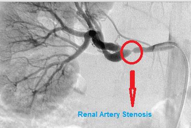
Fibromuscular Dysplasia
What is Fibromuscular Dysplasia?
Fibromuscular dysplasia can involve the leg arteries or rarely the arteries in other areas of the body. Usually it involves many arteries in the body.
The walls of the arteries are made up of three layers:
- Tunica intima or the innermost layer
- Tunica media or the middle layer
- Adventitia or the outermost layer
Fibromuscular dysplasia is classified based on the involved arterial layer and the lesions’ structure. Based on the classification of fibromuscular dysplasia, the narrowing of the artery is brought about by excessive fibers or muscle parts of the wall of the arteries.
Despite the fact that the classification of fibromuscular dysplasia can only be diagnosed 100% by examining the arterial wall under a microscope following a biopsy or surgery this is unusually done.
Generally it is likely to diagnose the classification of fibromuscular dysplasia depending on the form of the arteries involved in the dye angiogram examination.
Types
Here are the five classification of fibromuscular dysplasia, arranged from the most typical to the least:
1. Medial fibroplasia
75 to 80% of the cases of fibromuscular dysplasia are this classification
involves the middle layer (tunica media)
described by parts of fibrous lesions with aneurysms (bulgy parts)
looks like beads on the string in the dye angiogram (hallmark sign)
2. Intimal fibroplasia
occurs in lower than 10% of all the cases of fibromuscular dysplasia
brought about by fibrous tissue deposits surrounding the innermost layer (tunica intima) of the artery
without beads, smooth, concentric and narrowed appearance in the dye angiogram (hallmark sign)
3. Perimedial fibroplasia
lower than 10% of fibromuscular dysplasia are this classification
huge fibrous tissue deposits found in the adventitia or the outermost layer
irregular and thickened walls of the artery
beads that have a small diameter rather than normal artery (hallmark sign)
intensifies the probability of full obstruction of involved arteries
4. Medial hyperplasia
1 to 2% of lesions are under this classification
brought about by over formation of the muscular cells
no fibrous tissue deposits
the appearance is the same with intimal fibroplasia in the angiogram test
5. Periarterial hyperplasia
unusual less than 1% of the cases are in this classification
brought about by widening of the adventitia
the fibrous deposits spread around the fatty layers
defined by swelling of the artery
Symptoms & Signs
The majority of the people suffering from fibromuscular dysplasia are asymptomatic; however, symptoms can happen if the narrowing is serious enough to obstruct blood circulation on the involved artery.
Symptoms of moderate case of fibromuscular dysplasia in the carotid artery:
- headaches
- tinnitus (ringing in the ears)
- lightheadedness
Symptoms of advanced case of fibromuscular dysplasia in the carotid artery:
- stroke
- TIA or transient ischemic attack
Symptoms of carotid dissection (tear in the artery)
- headache
- acute neck pain
- stroke in advanced cases
- transient ischemic attack in advanced cases
Symptoms of fibromuscular dysplasia in the arteries in the kidney
- elevated blood pressure
- renal insufficiency
- commonly does not lead to renal failure
Causes
The exact cause of fibromuscular dysplasia is not yet known. This condition is most probably caused by many underlying reasons. Some of the reasons that may have a role involve:
Hormonal imbalances
Fibromuscular dysplasia happen mostly in women
Heredity
10% of all the cases are inherited. It may also be brought about by genetic defects affecting the arteries.
Trauma or stress
Trauma or stress especially on the walls of the arteries
Lack of oxygen to the wall of the blood vessel
This happen when the capillaries in the walls of the arteries that provide oxygen get obstructed by fibrous lesions.
Diagnosis
Diagnosis of Fibromuscular dysplasia is done with blood vessel studies. The below are common investigations to diagnose fibromuscular dysplasia
- Duplex ultrasound
- Angiography
- CT Angiography
- Magnetic Resonance Angiography

Treatment
The following are the treatments for fibromuscular dysplasia:
Medications
When fibromuscular dysplasia does not have any symptoms, it is typically benign and does not need management. For these people, the doctor may give an anti-platelet drug to avoid blood clots. The anti-platelet drug may be given or your doctor may advise that you use aspirin regularly.
People suffering from high blood pressure brought about by fibromuscular dysplasia may be prescribed with anti-hypertensive medications, especially ACE inhibitors.
Know and Control Risk Factors
Risk factors like hypertension, diabetes and elevated cholesterol should be assessed and controlled. People suffering from these problems must undergo CT scan, MRI or duplex ultrasound regularly to monitor the advancement of the disease. This is especially necessary when an aneurysm is confirmed.
Angioplasty
In special cases, percutaneous angioplasty of the arteries of the liver is advised. Same with the method applied to control obstruction in the cardiac arteries, renal angioplasty includes placing of a balloon catheter in the artery at the area of the narrowing or obstruction. The catheter is directed inside the blood vessel with the help of an X-ray. The balloon will be inflated to open the artery and then the balloon catheter is removed.
Putting a stent or a small tube at the area of the obstruction has not been tested to enhance the effectiveness of angioplasty and does not intensify the outcomes. Usually the stent is only used when angioplasty alone is not enough to enhance the blood circulation.
Angioplasty is also advised to those people suffering from fibromuscular dysplasia of the internal carotid artery who suffer from stoke or transient ischemic attack. Stenting may be needed in unusual cases, when people suffering from fibromuscular dysplasia have had vertebral or carotid artery dissection or aneurysm.
Surgery
Reconstructive surgery may be advised for people suffering from complicated fibromuscular dysplasia of the arteries of the kidney or suffering from an aneurysm of the internal carotid or vertebral arteries. Surgery will be based upon the area or the extent of the condition, even though usually it includes removal or bypassed of the involved part of the artery to recover the normal blood circulation.
Prognosis
At present there is no permanent cure for fibromuscular dysplasia. Medicines and angioplasty can lessen the probability of stroke. In unusual cases, aneurysms associated with fibromuscular dysplasia can rupture causing bleeding inside the brain that can lead to stroke, irreversible brain damage or death.