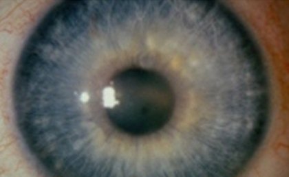
Fuchs Corneal Dystrophy – Symptoms, Treatment and Surgery
What is Fuchs Corneal Dystrophy?
Fuchs Dystrophy is a disease that involves the eye’s cornea. It is a condition that can be hereditary. The cornea is the transparent layer at the front of the eye that helps focus rays of light. It is very common that the disease includes both of the patient’s eyes and a little more frequent in females than males.
Pathophysiology
So now that we know the definition of this disease, we now need to know how this disease happens. The endothelial cells or the cells that compose the posterior layer of the eye’s cornea start to weaken and later on this will result to improper functioning. And then these cells little by little gradually will not be able to compensate. The cornea will now become condensed with additional fluid. In the later stage, the manifestations like for instance poor vision and pain in the eye may develop.
The treatment will vary from patient to patient. Some patient can be treated effectively with the use of prescribed ointments and eye drops. However, some patient may need a surgical transplant of the cornea.

Symptoms & Signs
Some physicians may already perceive early signs of Fuchs’ Endothelial Dystrophy in 30 years old and 40 years old patients. Nevertheless most patients are asymptomatic until they reach the age of 50 and 60, in which they start to manifest the symptoms.
Signs and symptoms of Fuchs’ Dystrophy include the following:
- It commonly involves both eyes
- Poor/Blurred vision
- The poor vision upon arousing may slowly improve as the day went by
Additional types of visual impairment include:
i. Vision may appear distorted
ii. Light sensitivity
iii. Poor night vision
iv. Halos around lights may be seen
v. Eye pain (generalized in nature)
vi. Epithelial blisters or tiny blisters that are painful on the patient’s cornea related to additional fluid inside the cornea
vii. Cloudy or hazy cornea
Blindness may occur in the later stage
It is advisable that if you have some of these manifestations, and particularly if they get really bad after a while, see your ophthalmologist or optometrist (eye doctor in layman’s term) for consultation. However, if signs and symptoms have appeared in a rapid manner, you have to call or see an ophthalmologist right away.
Treatment of Fuchs Corneal Dystrophy
As I have mentioned previously in this article, Fuch’s dystrophy can be treated effectively by simple means using prescribed eye-drops or ointments. These eye-drops and ointments help extract the extra fluid out of the patient’s cornea and these will eventually relieve symptoms of Fuch’s dystrophy.
However, if sores start to appear on the cornea and if these sores are very painful, the following can be prescribed:
i. Soft contact lenses
ii. Surgery
These two will help form the flaps above the sores that may aid in reducing the pain.
Corneal transplant is the mere cure for Fuch’s dystrophy. In fact, one of the chief reasons for corneal transplant in the U. S. is because of Fuch’s dystrophy. There is what they called Deep lamellar keratoplasty or DLK that is considered a substitute to a conventional transplant. DLK is a procedure wherein simply the cornea’s deep layers are restored with the tissue coming from the donor of the cornea. If you’re wondering if the procedure requires stitches, fortunately it does not. The good news is the time for convalescence is faster and there are few problems after the procedure.
The expected prognosis is that Fuchs’ dystrophy will get worse eventually without a transplant of the cornea. A patient unfortunately may lose his sight or he may have pain that is very severe and a very blurred vision in the later stage.
Usually a cataract surgery may aggravate mild cases of Fuch’s dystrophy. Therefore, a cataract surgeon should weigh the risk and dangers and he may change the procedure or the time of the scheduled cataract surgery.
The common complications include:
- Sensitivity to light
- Blindness that can mild to severe
- Pain that can be more rigorous and regular as the disease proceeds to its later stage
Remember to always call your health care provider if you have one of the following:
- Pain in the eye
- Light sensitivity
- You can feel that there is something inside your eye, but when you take a look there is really nothing there
- Halos around lights and cloudy vision
- A vision that is worsening over time
Surgery for Fuchs Corneal Dystrophy
It is typical that in the early stages of Fuchs’ dystrophy, no management is necessary. However, in some patients the disease may become too severe, wherein swelling of the cornea may begin. For these cases, drops of saltwater and ointment made of salt may aid in extracting the extra fluid out of the cornea and later help for the recovery of the patient’s vision. Sometimes using a hair dryer to momentarily dry the eyes can also be useful. Though the outcomes of these interventions may only be for short-term relief.
Surgery will become the last resort when the vision of the patient drastically becomes worse. Nowadays there are two known surgical methods that may be used. These are Descemets Stripping Endothelial Keratoplasty or DSEK and Penetrating Keratoplasty or PK.
First let’s discuss the Descemets Stripping Endothelial Keratoplasty or DSEK:
- In DSEK the lining in the cornea’s epithelium will be removed and substituted with a disc of the donor’s endothelial cells.
- The substitution of the faulty cells of endothelium will permit the cornea to once more become transparent by reestablishing the endothelial fluid pumping ability that was lost.
- It is done through incisions that are very small and because of that the healing time or recovery is very fast.
- The cornea will now then be greatly normal by four to six weeks time after the surgery.
- However there are some occasions wherein the donated endothelial cells will become dislocated within the cornea. When this happens it should be immediately repositioned through surgery or if it does not work it will need to be replaced completely. That’s all you need to know about DSEK. Now let’s talk about Penetrating Keratoplasty or PK:
- This is commonly used in severe cases wherein there’s already corneal swelling or corneal scarring.
- During the procedure, the surgeon will need to remove the whole cornea and then he will need to replace it with a complete cornea from a donor.
- The sutures which are similar to a hair of a person will be used to secure the donor tissue in the patients’ cornea.
- For quite a few months, the sutures will remain in place and will be gradually removed over a year’s course.
- This took a longer recovery but considered still as the better option for visual cure rather than DSEK.
Pictures
