Archive for March, 2012

Renal Tubular Acidosis – Symptoms, Diagnosis and Treatment

Mar 31 2012 Published by under Diseases & Conditions

What is Renal Tubular Acidosis?

Renal tubular acidosis or RTA is a condition wherein there is an accumulation of acids in the body because of failure of the kidneys to perform its function in acid-base balance, specifically to excrete acids into the urine. When blood passes through the kidneys, it is filtered to make the blood clean. The filtrate passes again into the renal tubules for the exchange of acids, salt, and other solutes before urine drains into the bladder to be eliminated. The metabolic acidosis caused by renal tubular acidosis may be caused either by failures in the renal tubules such as in the proximal and distal renal tubules. When there is insufficient reabsorption of bicarbonate ions (alkaline) in the proximal tubule or there is inadequate excretion of hydrogen ions (acid) in the distal tubule, then acidosis may occur. Renal tubular acidosis involves the insufficient urinary acidification in well-functioning kidneys as opposed to the metabolic acidosis that occursin renal insufficiency.

Acidosis occurs when the pH of the blood is below 7.35. It was first described in children by Lightwood and Butler et al. in 1935 and 1936 respectively. RTA in adults on the other hand was first described by Baines et al. in 1945.

Metabolism in the body generates acids as byproducts. Presence of acids in the blood is normal, but an excess of it can lead to disruption of body functions. Acidosis is prevented by the kidneys by excreting acids into the urine and reabsorbing bases or alkaline back into the blood to maintain a normal pH.
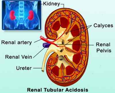
renal tubular acidosis

Source – medindia.net

There are different types of renal tubular acidosis depending on the location of failure. These also have varied causes and manifestations. These include:

a. Distal renal Tubular Acidosis or Type 1 RTA

Distal RTA or classical RTA involves the failure of the distal renal tubules to secrete acid in the urine. The inability to excrete acids leads to a non-acidic urine of >5.3. Acids are primarily excreted through the urine. When this is impaired excretion, acidemia or acidosis occurs.

The inability of the cells to excrete hydrogen ions also leads to non-reabsorption of potassium leading to hypokalemia and other electrolyte imbalances.

Causes of distal RTA

  • Inheritance – Defects in the distal tubules were studied to be inherited because of abnormal genes present.
  • Systemic Autoimmune disorders – Diseases such as Sjögren’s syndrome and systemic lupus erythematosus also destroy the distal tubules leading to impairment of the functioning of the area.
  • Diseases that cause abnormal calcium deposits in the kidneys – Diseases such as sickle cell anemia, hyperthyroidism, hyperparathyroidism, primary biliary cirrhosis, chronic active hepatitis, analgesic nephropathy, kidney rejection, obstructive uropathy and urinary tract infections cause calcium to build up on the nephrons causing impairment of distal tubular functions.

Symptoms of distal RTA

  • Normal anion gap metabolic acidosis
  • Urinary stones as a result of alkaline urine
  • Hypokalemia
  • Nephrocalcinosis
  • Bone demineralization as a result of hypophosphatemia (rickets in children and osteomalacia in adults)

b. Proximal Renal Tubular Acidosis or Type 2 RTA

Proximal RTA is primarily caused by inability of the proximal renal tubules to reabsorb filtered bicarbonate ions from the urine. The excretion of bicarbonate ions leads to wasting and subsequently, acidemia because there is insufficient alkaline in the blood to neutralize or buffer the acids. The acidosis in proximal RTA is less severe than distal RTA because the kidneys still secrete acids into the urine.

Causes of Proximal RTA

  • Fanconi’s syndrome – This is a condition where there is generalized dysfunction of the proximal renal tubules. It is associated with phosphaturia, hypokalemia, glycosuria, aminoaciduria, uricosuria and proteinuria because of the inability of the proximal tubules to reabsorb these chemicals, including bicarbonate ions. Because of phosphate wasting and Vitamin D deficiency, there is bone demineralization (rickets or osteomalacia)
  • Cystinosis – Cystinosis is an inherited disorder that disrupts the normal breakdown and use of nutrients. Cystine crystals are deposited in the bones and cause non-reabsorption of bicarbonate ions. Other diseases include Wlison’s disease and fructose intolerance.
  • Chemotherapy – Ifosfamide, a chemotherapeutic drug disrupts the functioning of the proximal tubules leading to acidosis.

Symptoms of proximal RTA

Excretion of bicarbonate ions leads to non-reabsorption of other electrolytes giving rise to the following symptoms:

  • Hypokalemia (low potassium in the blood)
  • Phosphatemia
  • Urine pH of 5.5 (alkaline)
  • Reduced excretion of Hydrogen ions
  • Impaired reabsorption of bicarbonate ions
  • Impaired exchange of sodium, potassium and chloride in the distal tibule as a result of aldosterone resistance

Symptoms of Renal Tubular Acidosis

Aside from the specific symptoms discussed earlier, the primary manifestation of RTA is on the level of potassium in the blood. Distal and proximal RTA result in hypokalemia. Potassium is essential for the contractility of the heart and nerve impulse transmissions. A low or high potassium levels result in the following symptoms:

  • Weakness, fatigue
  • Paralysis
  • Dysrhythmias or irregular heart beat
  • Anorexia, vomiting
  • Abdominal distention
  • Leg cramps
  • Postural hypotension
  • Shallow respirations
  • Lethargy and confusion

Causes of Renal Tubular Acidosis

Specific causes of renal tubular acidosis according to type were discussed earlier. General classifications of causes for all types of RTA include:

  • Genetics
  • Autoimmune diseases
  • Hypercalcinosis (calcium deposit build up)
  • Drugs or medications
  • Other renal disorders

Diagnosis of Renal Tubular Acidosis

RTA diagnosis includes the following tests and exams:

  • Acid-base balance examinations – Arterial blood gas studies help in determining presence of acidosis through collection of blood samples on the artery. Urine sample is also taken to check for urine acidity. A high blood acidity and low urine acidity indicates renal tubular acidosis.
  • Electrolyte tests – Checking the electrolytes especially the potassium is essential to check presence of hypokalemia or hyperkalemia. Abnormal potassium levels are life-threatening because it cause changes in the contractility of the heart which may lead to cardiac arrest. Other electrolytes include sodium, chloride and phosphate. This identifies the type of RTA a patient has.

Treatment of Renal Tubular Acidosis

Treatment of acidosis focuses on neutralizing the pH of the blood. Electrolyte imbalances are also corrected. Treatment approach includes the following:

  • Administration of Alkali – Sodium bicarbonate or sodium citrate and citric acid (Shohl’s solution) is administered daily in divided doses to increase bicarbonate levels and normalize the pH of the blood. However, administration of bicarbonates also leads to further hypokalemia so potassium replacement should be instituted. Treating acidosis prevents renal failure and calcium stone formation. Alkali therapy also reverses problems on sodium, chloride and phosphate abnormalities.
  • Potassium Replacement – Potassium is given to correct hypokalemia. In cases of type 4 RTA with hyperkalemia, diuretics such as furosemide may be given to excrete excess potassium levels in the blood.
  • Hydrochlorotiazide Diuretics – Hydrochlorothiazide are special kid of diuretics that allows reabsorption of potassium in the tubules leading to increase in potassium levels during bicarbonate therapy because administration of bicarbonate leads to more excretion in the urine of bicarbonate ions and potassium.
  • Phosphate and Vitamin D therapy – In cases of Fanconi’s syndrome in proximal RTA, Vitamin D and phosphate should be replaced to prevent bone deformities such as osteomalacia and rickets

Prompt management of RTA prevents further complications and renal failure.

Complications of Renal Tubular Acidosis

Renal tubular acidosis results in the following complications as a result of interplay of electrolyte imbalance, calcium formation and Vitamin D deficiency:

  • Bone disease. Hypophosphatemia leads to bone demineralization because phosphate is responsible for calcium resorption in the bones. Inability to maintain calcium in the bones leads to bone breakage and calcium stone formation in other parts of the body.
  • Growth retardation. The occurrence of rickets and osteomalacia result in retarded growth.
  • Kidney stones. Calcium deposits as a result of alkaline urine and hypophosphatemia eventually lead to kidney stone formation which may potentially lead to renal failure.
  • Renal Failure. This is the result of RTA when treatment is not done to correct the condition.

Prevention of Renal Tubular Acidosis

Prevention of RTA includes measures to prevent the causes. It is essential to maintain good hydration by increasing fluid intake to prevent stasis of byproducts in the kidneys. Avoiding highs salt diet is also essential to prevent overworking the kidneys to regulate sodium levels. Ensure adequate exercise to prevent calcium stasis and stone formation. Genetics, inheritance and certain autoimmune diseases that lead to RTA cannot be prevented so supportive managements should be implemented to maintain healthy kidneys.
No responses yet

Lump on Back of Neck and Head – Causes and Treatment

Mar 29 2012 Published by under Diseases & Conditions

What is Lump on Back?

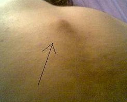
According to research, the reported cases of patients having lumps on the back are very minimal. In fact, this condition is of very rare occurrence. Most often of the cases reported are not of a serious kind. At times, you might even find that a lump is present and within days, it will disappear even without medical interventions. Hence, this kind of lump is part of the not serious category. Hence, you need not be alarmed. However, there are painful or discolored lumps that don’t disappear quickly and will persist for a longer duration. You need to let the physician evaluate this lumps as soon as possible to be able to know the reason behind it.

Lump on Back

Picture 1 – Lump on Back

Lump on Back Causes

The lumps on the back can be either be found in the neck or head part. As we go along, the possible reasons or the etiologies will be discussed.

Lump on Back of Neck Causes

There are two primary reasons why there are lumps in the neck. The reasons are broken down into due to a swollen lymph nodes and salivary gland enlargement.

Swollen Lymph Node. Under the swollen lymph node category, the lumps of the neck might be due to bacteria, cancer, and thyroid, viral and allergic reaction in origin.

Salivary Gland Enlargement. Under the Salivary Gland Enlargement is the infection, mumps, salivary gland tumor and stone in salivary duct which all leads to having a manifestation of lumps in the neck region.

It may be Bacterial in origin which may be due to:

  • Atypical Mycobacterium. It is a type of bacteria that also affects the HIV patients who has a CD4 count of 50 or less. This kind of bacteria when shown positive in a person will manifest swelling and painless lymph nodes in the neck area.
  • Cat Scratch Disease. This kind of disease is due to the fact that a cat has scratched a person. After being scratch by a cat, within three to five days the signs will appear. One sign will be developing a red bump. It will later progress into non-itching papules and then will turn into crust after three days and will eventually heal with a scar. Three to four weeks after being scratched, the person will manifest solitary lymphadenopathy or having enlarged lymph nodes. This will be painful but rest assured that it will resolve after four months maximum.
  • Pharyngitis. This disease condition is an inflammation of the pharynx or the tonsils. It is a viral and self limiting disease. When it becomes severe, it may lead to having tender lymph node enlargement on both sides of the neck.
  • Peritonsillar Abcess or Strep Throat. It is an extension of the infection of the tonsils which is beyond the capsule. It also has abscess formation which is located behind or above the tonsil region. It can cause swelling or lump in the neck which is tender to touch.
  • Tuberculosis. This disease condition usually affects the lung. Systemically the tuberculosis can affect any part of the human body. If this condition is present in the nodes of the neck, it will result to a large lump having an irregular margin.
  • Tonsillitis. It is an infection of one or both the tonsils. When a person has this disease condition, it will manifest swelling or a lump in the neck area.

It may be due to Cancer such as:

  • Hodgkin’s and Non- Hodgkin’s Lymphoma. These are known tumors of the immune system. Both disease condition share similar symptom that the patient manifests, which is the swelling or the lump found in the neck. The neck has a region where lymph nodes can be found. When these lymph nodes are infected, it enlarges and causes lumps or swelling in the neck.
  • Leukemia. In leukemia, the white blood increases in number. Hence, this might signify the start of the inflammation process. One of the sign of inflammation process is having lumps or swelling in areas of the body. Some of these lumps might be found in the neck region.
  • Mouth Cancer. Oral or mouth cancer is a tumor located in the mouth. Once the tumor progresses, it will manifest sore or lumps in the neck area especially if it is in the advanced state.

It may also be due to Thyroid problems such as:

  • Thyroid Cancer. Lumps of the neck may be due to Thyroid cancer. It is an abnormal thyroid tissue with uncontrollable proliferation and it has a tendency of invading the lymph nodes that are found in the neck. Hence, causing the lumps which when felt will feel non-tender and hard.
  • Goiter. Enlargement of the thyroid gland is called as Goiter. When one has this disease condition, he or she will manifest lump or swelling of the neck region.
  • Grave’s Disease. When one has Grave’s Disease, will manifest similar to the Goiter patients. Compared to goiter, this disease is autoimmune and is primarily due to thyroid gland over activity.

It may be Viral in origin which may be associated with:

  • AIDS. The AIDS which stands for Acquired Immune Deficiency Syndrome is the final and last stage of HIV disorder. This will target and damage the immune system per se. Since it will damage the immune system, it will lead to inflammatory process. One inflammatory process is swelling of the lymph gland which will manifest lumps in the neck.
  • HIV disease. It is a condition that is primarily caused by Human Immunodeficiency Virus. This virus will cause injury to the immune system which will make it harder for the body to fight opportunistic infections. It will lead to the person having swelling or lump of the neck.
  • Infectious Mononucleosis. This disease condition is caused by a member of the herpes virus family. It will lead to the manifestation of swollen of lymph nodes which will lead to lump of the neck.

Other causes may be due to Allergic Reaction

This allergic reaction will target the inflammatory process which will lead to having a swelling or lump of the neck, which is one of the symptoms under the inflammatory process.

Lump on Back of Head Causes

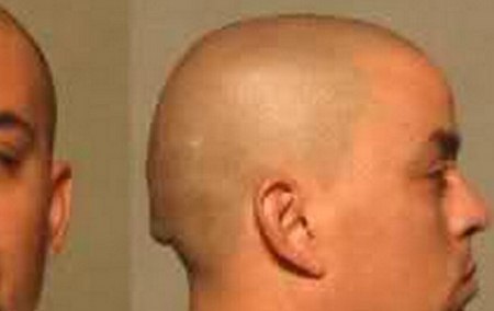
In some instances, it is difficult to tell that you have a lump in your back of the head or head lumps for that matter, with just one look. The lumps on the back of your head may be due to the following:

Lump on back of head

Picture 2 – Lump on back of head

Dermatitis. It is when the skin of the scalp found in the head becomes inflamed and itchy. It can then lead to weeping of fluid that when it dries, it will form in to crust leading to lumps in the head.

Skin Cancer. Some of the skin cancer may appear on the head particularly in the scalp region. When it appears in the scalp it can be described as a growth like a bump or lump in the head.

Cysts. Epidermal or sebaceous cysts are caused by a duct located at the basement of the hair shaft that was clogged. It will also lead to having lumps on the head portion.

Hive. Hives are due to allergic reaction. It will be manifested in large red bumps or lumps.

Lump on Back Treatment

Lump on Back of Neck Treatment

  • Pharmacological Intervention. When the etiology of the lumps of the back of the neck is due to bacteria or viral cause, antibiotics may be given. You may also give anti-inflammatory medications to reduce swelling.
  • Surgical Intervention. When despite pharmacological intervention has been done, yet the lump is not resolved or treated. Then, the last option left would be its Surgical Intervention.

Lump on Back of Head Treatment

  • Medication. When the etiology of the lump of the back of the neck is due to dermatitis, you can use medical shampoos out in the market such as ketoconazole.
  • Surgery. If the cause of the lump of the back of the head is due to cyst, malignant or benign in nature. Then it is necessary to remove it as soon as possible to prevent the spread of the cyst.

No responses yet

9 Tips to Lose 5 Pounds in a Week

Mar 28 2012 Published by under General Health

Want to lose extra pounds fast? Have you tried a lot of slimming pills and diet techniques, but offered no solution for your weight? Losing weight has never been easy to those who wanted it so much. However, there are some techniques you can do to shed off that extra 5 pounds for an upcoming event or date.

Lose 5 Pounds in a Week

Here is a proven weight loss remedy that can make you lose pounds in just a week. These are used by athletes who need to shed off weight for an upcoming weigh in and also effective for normal individuals who want to lose weight.

lose 5 pounds in 1 week

Image Source – people.co.uk

1. Drink a lot of water

Although water increases the weight suddenly when you drink too much, drinking a lot will reduce your food intake because this will make you feel full. In addition, water also does not contain fats, carbohydrates, sodium and calories that other beverages contain. The kidneys has the ability to eliminate water in the body and the water you take in will just help you reduce calories, but not add to your weight. Water may also help detoxify the body.

2. Eat whole bread, grains and pasta

Whole grains make you feel full fast without even eating too much. In contrast, white bread and pastas makes you eat more because they are digested faster than the unrefined grains. White bread and pasta also have more empty calories that quickly add up to your weight. But if you can eliminate eating these carbohydrates in a day, then it is better.

3. Indulge in fruits and vegetables

Who says you cannot eat fruits and vegetables more in a day? Nutritionists and the food pyramid suggest that among all the food groups, vegetables and fruits should be taken in larger quantities. Fruits and vegetables make you full without those unwanted calories and fats. Substitute your usual breakfast with a piece of banana or apple. Eat vegetable salad instead of a regular meal during lunch and make vegetable soups for dinner. Eating this kind of diet for a week will definitely cut those excess pounds. Just remember to include small portions of carbohydrates and proteins in your diet because these are still essential for energy production and cell repair.

4. Work out at least 30 minutes a day

Consider doing cardio exercises, lunges, pushups and abdominal curls. These types of exercises burn more calories than any type of exercises. Instead of using the treadmill, consider using the stationary bike because this significantly burns more calories.

5. Sleep 30 minutes to one hour longer than you used to

Having a good sleep enhances the building of muscles. When people sleep, the body builds muscles and improves the muscle tone. Having quality sleep also makes you feel better inside and out.

6. Discipline

The key to a successful weight loss program is discipline. Before you start your week losing weight, try to condition your mind. You can post messages on the fridge, your room, bathroom and other places to remind you of your goal to lose weight.

7. Choose slimming clothes

This is an instant way of creating an illusion that you have shed extra ten pounds. Go for dark, monochromatic shades, V-neck shirts, A-line skirts, clothes with vertical prints, thin garments, long pants, properly fitting underwear, platform wedges and big accessories. These techniques create an instant illusion that you are thin even if you have not shed any pounds.

8. Engage in a nightly sex.

Yes, this is very effective in burning up calories. In fact, sex burns up to 200 calories in 30 minutes. It is also better if you perform a lot during sex to increase the rate of calorie burning. It does not only enhance calorie break down, but also increases the over-all well-being and confidence.

9. Cut-out fats and sodium

Refrain from eating junk foods, fried dishes, and dining out in fast foods. Fats and sodium increases the weight and also are not healthy when taken in excessive amounts. Sacrifice not eating these items and you can achieve your goals.
When these suggestions are applied continuously, it will just not temporarily help you lose 5 pounds in a week, but continue to help you achieve a more slender body.

No responses yet

Slipped Disc – Symptoms, Treatment and Surgery

Mar 27 2012 Published by under Diseases & Conditions

What is Slipped Disc?

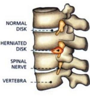
Slipped disc happens when the intervertebral disc ruptures or herniates. The herniation or rupture causes the low back pain, sensory changes in the lower limbs and even immobility. Slipped disc is a common term but this condition is more appropriately known as herniated nucleus pulposus.

slipped disc and normal disc

Image source – drapbandodkar.com

The Spine

In slipped disc or herniated nucleus pulposus, it is the spine that is primarily affected. Our spine moves like a spring when we are lifting and moving. The spine adjusts and disperses the weight by compression and expansion in situations where we lift heavy loads and when we do vigorous activities. The spring like action of the spine distributes the weight evenly, preventing the spine and its surrounding structures to be compressed. Our spine is pushed to the limits when we lift enormous loads and when we are overworked.

What Happens With Slipped Disc?

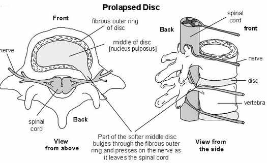
The human is made up of individual intervertebral discs; these intervertebral discs have two compositions, an outer ring made up of connective fibrous tissue, and an inner core that is gelatin like. The outer ring is hard and it protects the soft gelatin like core, this core is called the nucleus pulposus (hence the term herniated nucleus pulposus).

When the spine experiences heavy workloads (as in heavy lifting), the nucleus pulposus absorbs and distributes the load all throughout the intervertebral disc. When the load is too much, herniated nucleus pulposus happens and the soft central structure comes out of the fibrous ring. This herniation causes nerve compression, pain and inflammation.

Slipped Disc pathophysiology

Image Source – patient.co.uk

Symptoms and Signs of Slipped Disc

The following symptoms and manifestation may indicate you have a slipped disc of spinal disc herniation:

  • Mild to severe neck and low back pain
  • Pain that radiates to other areas such as the feet, knees or thighs
  • Pain in the shoulder, clavicles, or jaw
  • Tingling sensation in the upper or lower limbs
  • Weakness of the muscles (in the back or neck)
  • Paresthesia
  • Possible paralysis
  • Hypoactive or hyperactive reflexes
  • Sciatica due to the possible irritation of the sciatic nerve
  • Pulsating and/or recurrent pain
  • Possible loss of bladder, bowel and sexual function

Signs and symptoms can be mild to severe depending on the area affected by the herniation. Some individuals may have no symptoms at all while some may have more pronounced manifestations.

Symptoms are usually experienced on one side of the body (unilateral), however if the herniation is large and it happens on the cauda equina on the lumbar region, then symptoms may be felt on both sides of the body. If loss of bladder and bowel control is present, the individual must seek immediate medical attention.

Causes of Slipped Disc

The following can cause or lead to slipped disc or herniated nucleus pulposus.

  1. Wear and tear of the spine
  2. Degeneration to the spine brought about by aging or other underlying conditions
  3. Improper body mechanics while lifting
  4. Lifting of heavy weights
  5. Trauma
  6. Poor posture
  7. In some cases mutation of the genes responsible for the strength of the nucleus pulposus

Risk Factors of Slipped Disc

Certain individuals with certain conditions are at a greater risk of experiencing a herniated disc. Individuals at risk include the following:

  • People who do heavy lifting such as construction workers, heavy machineries workers, and even doctors and nurses
  • Prolonged sitting
  • Athletes or weight lifters
  • Obese or overweight individuals
  • Smokers
  • Workers with strenuous activities

Slipped Disc in the Neck

Slipped disc that happens in the neck is called cervical herniated nucleus pulposus. This could be a delicate condition since the neck or the cervical area is the highest region of the spine. Any damage to this area means significant loss of function of the areas below it.

This means significant nerve damage possibly brought about by a herniation can lead to paralysis or an impaired function in the parts of the body from the neck down.

Signs and Symptoms of Cervical Disc Herniation

  • Mild to severe pain in the neck
  • Pain while extending and flexing the neck
  • Pain sneezing, straining or coughing
  • Pain that radiates to the upper arm to the wrists and even fingers
  • Possible weakness of the arm muscles
  • Weak hand grip
  • Numbness or tingling sensation in the arms and hands

Slipped Disc in the Lumbar Area

Disc hernia that occurs in the lumbar area is one of the most common types of disc herniations. Lumbar slipped disc is also one of the most common causes of lower back pain complaints in adults.

Signs and Symptoms of Lumbar Slipped Disc

  • Low back pain which can be chronic or acute
  • Sensory changes such as tingling and numbness of the lower extremities
  • Mild to severe pain radiating to the buttocks, thighs, and feet
  • Weakness of the lower extremities
  • Possible paralysis

Danger symptom: Cauda equine syndrome- this involves loss of bowel and bladder control.

Diagnosis

Slipped disc can be diagnosed by implementing the following procedures:

Imaging studies such as:

  1. X-rays
  2. Computed Tomography (CT Scan)
  3. Magnetic Resonance Imaging or MRI
  4. Electromyogram and Nerve conduction studies (EMG-NCS) to diagnose nerve damage
  5. Physical examination through the straight leg raise test

Treatment

Slipped disc treatment involves both non-surgical and surgical interventions. In most cases the disc hernia heals within six weeks so surgery is not done. But for cases where there is a large protrusion and nerve damage becomes a complication, surgical measures are done.

Non-Surgical Treatment

NSAIDS and Cortisone Injection

For mild cases treatment usually involves medications such as NSAIDS or Non-steroidal Anti-inflammatories. The medication treats pain and inflammation.

NSAIDS are usually taken orally but for individuals who use the medication for a long time, another mode of treatment is given. This is to prevent cardiovascular and gastrointestinal toxicity. For these individuals, cortisone injections are given to relieve the pain and other symptoms.

Ergonomics and proper body mechanics

Designing equipment and work environment in ways that it prevents back injuries and further slipped discs are advised treatments. This usually involves the use of assistive devices for lifting weight and preventing back strain. Following proper body mechanics is also an important. Lifting should follow one’s center of gravity this keeps the spine straight and disperses the weight evenly.

  • Use of supportive devices on the back
  • Control of weight
  • Traction therapy
  • Intake of Oral Steroids
  • Epidural injection of cortisone

Physical Therapy

Physical therapy is usually done with other therapies. Physical therapy focuses on relieving the compression through exercise and the use of tractions. Physical therapy also prevents the possible complications.

Surgical Measures

  • Chemonucleolysis– surgical procedure that dissolves the herniated nucleus pulposus
  • Discectomy/ microdiscectomy- the removal of the protrusion to aid in the compression of the nerves.
  • Laminectomy– spinal lamina removal that allows ample space for the compressed nerves and adjacent structures. It is usually done when a spinal stenosis is suspected.
  • Hemilaminectomy– removal of a portion of the lamina
  • Artificial disc replacement- this surgical procedure is done to individuals with a herniated disc due to degeneration.
  • Nucleoplasty– nucleus pulposus repair
  • Tessys method (transforaminal endoscopic surgical system) – this is a less invasive surgical procedure in treating disc hernia. It involves the removal of the herniated disc through small incisions in the skin where an endoscope will be inserted to view the site affected. In this type of slipped disc treatment, local anesthesia is used instead of a general anesthesia, and damage to the adjacent tissues is minimal.

Complications

When herniated nucleus pulposus is not treated appropriately, the following complication may happen:

  • Cauda equine syndrome which involves an impaired bowel and bladder control
  • Permanent injury to the nerves
  • Paralysis and total loss of sensation to the affected limbs
  • Chronic pain

No responses yet

Slapped Cheek Syndrome – Pictures, Symptoms, Treatment

Mar 25 2012 Published by under Diseases & Conditions

What is Slapped Cheek Syndrome?

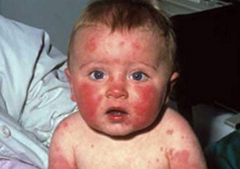
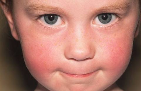
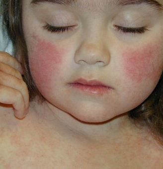
Slapped Cheek Syndrome is sometimes called as parvovirus infection, erythema infectiosum, Latin word which means infectious rash, or fifth disease. It is famously called as the fifth disease because of the fact that it is considered before as the 5th common disease of children. It is called as the slapped cheek syndrome because it manifests the child having bright red cheeks, as if they were actually slapped in their cheeks. The children, around the ages of 4 to 12 years old, are the ones often affected with this disease condition. It can happen, however, in any age. It is a viral form of disease condition. When one has this kind of syndrome, its incubation period is usually 4 to 20 days before the appearance of the rashes. When the rash appears, the syndrome is normally no longer infectious.

Symptoms and Signs

When a person has slapped cheek syndrome, he or she is considered infectious during the 4 to 20 days before the rashes develops. Hence, the signs and symptoms that one should watch out are the following:

  • Fatigue
  • Sore throat
  • Runny nose
  • Stuffy nose
  • Slight elevation of the person’s body temperature
  • Joint pain
  • Light pink rash
  • Neurological problems (rare)
  • Cardiovascular problems (rare)
  • Nausea
  • Diarrhea
  • Abdominal pain
  • Blotchy red appears on the person’s cheeks
  • Itchy
  • The rash spreads through the body and it takes 3 weeks to go away.
  • Headache
  • Irritability
  • General flu-like symptoms
  • Arthralgia or joint soreness
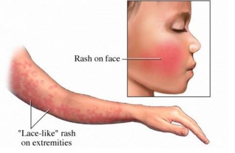
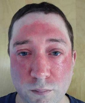
Slapped Cheek Syndrome (5th disease) - rash on face
Observe the rash on face in Slapped Cheek Syndrome

Causes and Risk Factors

The causative factor of slapped cheek syndrome is the parvovirus B19. However, there are parvovirus that is found in the cats and dogs. It is only the parvovirus B19 that is found commonly in human beings. The animals can’t be infected with the parvovirus that is associated with human beings and vice versa. The virus, itself, spreads from one person to another via airborne, direct contact, or saliva secretions of other persons.

Among the most common transmittable form of slapped cheek syndrome is through coughing, hand to hand contact, or sneezing. Those people living in a secluded, congested area are known to be risk of having this kind of syndrome. The more the immune system is compromised, as like children and sick persons, the greater the risk they have to acquire the slapped cheek syndrome.

Diagnosis

The physician usually diagnoses the slapped cheek syndrome by doing the following actions:

  • Physical Examination
  • Medical History
  • Blood test to check for the status of your immunity

Amongst the test mentioned, the blood test is the only diagnostic test that will confirm the slapped cheek syndrome. If however, the blood test shows that the patient is immune. It means that there is no possibility that the patient will be re-infected with the syndrome. Yet, if the blood test shows that there was a recent infection of the parvovirus, it will require further examinations to find out if there is a presence of complications that requires treatment.

Treatment

There is no exact medication for the slapped cheek syndrome. It only requires supportive kind of treatment. Supportive treatment may include the following:

  • Rest especially needed to prevent fatigue
  • Paracetamol or Acetaminophen will relieve the fever symptoms
  • Cold flannels that will relieve the hot cheek discomfort as it will lead to vascular constriction
  • Adequate fluid intake to prevent dehydration especially when the person is having diarrhea
  • Antihistamines to relieve the itchy symptoms
  • Moisturizing lotion to soothe the itchy feeling
  • NSAIDs to relieve pain

Complications

Aside from the risk mentioned above, the high risk group may be able to have complications such as:

  • Infection in Pregnancy. Pregnant women can be infected before and during their pregnancy. Their babies have won’t be harmed but about 5 percent may be able to develop severe form of anemia which will lead to still birth or miscarriage.
  • Persons with Weak Immunity. Persons who are sick are usually the ones who have weak immunity. They are the ones called immunocompromised persons. Once they have weak immune system, they are at risk for acquiring diseases easily. One of the diseases that they are at risk of having is the slapped cheek syndrome. They manifest the symptoms of having severe anemia, a not well feeling, and a high temperature or fever.
  • Persons having Hemolytic Blood Disorders. Persons with blood disorders such as thalassemia, sickle cell anemia, and hereditary spherocytosis will have decrease hemoglobin. The most common complications are that of having severe anemia which will lead to aplastic crisis. In severe case, it will lead to having heart failure.
  • Arthritis. It is a possible complication of the virus that causes this syndrome which affects the small joints in the feet and hands. Symptoms are the same with those of rheumatoid arthritis. This kind of complication occurs mostly in adults.

Prevention

As of the moment, there is no vaccination that is available in preventing the slapped cheek syndrome. Persons, who were once infected with the parvovirus B19, are already immune with this kind of disease condition.

  • Hand washing. In preventing the slapped cheek syndrome, the first and safest thing to do is to practice hygienic measures such as daily washing of hands before and after eating and going to the comfort room. This kind of act will reduce the spread of infection.
  • Avoid sharing of utensils. The sharing of utensils should be also avoided. Consult physician as soon as possible if you manifest the symptoms mentioned. Pregnant women, who were exposed, should consult their physicians as soon as possible to prevent further complications.
  • Keep the immune system healthy. You need to keep the immune system healthy by eating well, having a complete eight hours sleep and consuming vitamins to prevent acquiring the disease condition.

Pictures

 Slapped Cheek Syndrome pictures Slapped Cheek Syndrome images

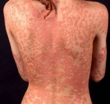
Slapped Cheek Syndrome pics rash on face Slapped Cheek Syndrome (5th disease)

No responses yet

Prurigo Nodularis – Treatment, Causes, Symptoms and Pictures

Mar 24 2012 Published by under Diseases & Conditions

What is Prurigo Nodularis?

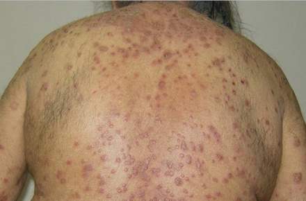
In layman’s terms, it means “bumps that are itchy”. It is a skin disease, which appears on the legs, arms and elsewhere in the body, which has a characteristic of nodules that are pruritic or itchy. It is also known as Hyde Prurigo Nodularis, PN, Lichen Simplex Chronicus, Picker Nodules, Lichen Corneus Obtusus, and Neurodermatitis Circumscripta in an atypical nodular form. The middle-aged to elderly persons are often the persons affected by this kind of skin disease. It is a chronic kind of inflammatory dermatosis or skin disease that results to papulonodular eruption. It has a characteristic of pruritus in a severe form, nodules and papules with ulcerations and excoriations with is because of too much scratching.

Prurigo Nodularis in women

Picture 1 – Prurigo Nodularis in women


Causes

The main etiology of why the Prurigo Nodularis exist in some persons is, up to this point, still unknown. Researchers found out that the following factors may induce the skin disease called Prurigo Nodularis, such of these factors include:

  • Fungal infections
  • Poor diet
  • Yeast infections
  • Toxin
  • Allergic disorders
  • Poor liver function
  • Psychiatric Illness such as delusion of parasitosis
  • Parasites
  • Thyroid disease
  • Linear IgA disease which is an autoimmune disease condition
  • Becker’s Nevus

It has also been associated and linked with the following disease conditions:

  • Hodgkin’s disease
  • Uremia
  • Polycythemia rubra vera
  • Thyroid disease
  • Cholestasis
  • HIV

Symptoms & Signs

When a person, at around 20 to 60 years old of age, is diagnosed with Prurigo Nodularis, he or she may manifest the following signs and symptoms:

  • Small red bump (early sign)
  • Brown, hard globular nodules
  • Bumps
  • Itching usually occurring at anytime but it most often occurs in the late evening and night

Diagnosis (Histology)

The best way to diagnose this kind of skin disease is done through:

  • Skin biopsy
  • Lesion biopsy which shows elevated eosinophils
  • Culture that is needed to rule out any infection especially staphylococcus infection which is linked to atopic dermatitis
  • Histopathology or Histology

Histology is the study of anatomy of the cell, which in this case is a human cell, under the microscope. Upon the histopathological exam, with the person diagnosed with this disease condition, will revealed orthokeratotic acanthosis and hyperkeratosis. With focal parakeratosis, under hyperkeratosis, there is an irregular acanthosis. The rete ridges are irregular and elongated with an infiltrate dense dermal that consists of eosinophils, histiocytes, neutrophils, and monocytes. The appearance of pseudoepitheliomatous or pseudocarcinomatous hyperplasia comes from a downward, irregular epidermis proliferation, severe papillomatosis and adnexal epithelial structures.

With the focused turned into the dermis’ papillary, there is an observed elevation of fibroblast that is multinucleated with thick fiber collagen bundles that are arranged to the surface, perpendicularly. There is also an observation of proliferation of the Schwann cells and nerve fibers. In some areas, they are vertically-oriented and dilated capillaries. Found at the surface, located in the interstitial spaces and around the vessels are moderately dense lymphocytes, isolated mast cells, eosinophilic granulocytes, macrophages, melanophages, dermal dendritic cells or hemosiderophages with erythrocytes that are extravasated. You can also find in patients diagnosed with atopic diathesis, eosinophilic granulocytes that are degranulated. Crusting seen around the margin with parakeratosis and exudates maybe found where there is a presence of excoriations or erosions and when there are neutrophils and plasma cells.

Differential Diagnosis

Other conditions that have similar appearances or the differential diagnosis with Prurigo Nodularis are the following conditions:

  • Impetigo
  • Insect Bites
  • Acute Febrile Neutrophilic Dermatosis
  • Aphthous Stomatitis
  • Bechet Disease
  • Atopic Dermatitis
  • Allergic contact Dermatitis
  • Lichen Ruber Verrucosus
  • Lupus erythematodes hypertropicus et profundus
  • Linear IgA Dermatosis
  • Dermatitis Heretiformis
  • Pemphigoid Nodularis
  • Bullous Pemphigoid
  • Chancroid
  • Chrug-Strauss Syndrome
  • Ecthyma
  • Lymphoma
  • Pseudolymphoma
  • Cutaneous metastases
  • Keratoacanthoma
  • Dermatofibroma
  • Ictus Reactions
  • Prurigo Pigmentosa
  • Actinic Prurigo
  • Polymorphous light eruption
  • Squamous Cell Carcinoma
  • Ecthyma Gangrenosum
  • Herpes Simplex
  • Wegrener Granulomatosis
  • Verrucuous carcinoma
  • Hypersensitivity Vasculitis or Leukocytoclastic Vasculitis
  • Venous Insufficiency
  • Sporotrichosis

Treatment

It is a challenge when the person will undergo treatment for Prurigo Nodularis. The treatment goal for this skin disease condition are to develop an individualized treatment scheme and topical and the usage of causal, systemic and topical therapies

Causal Therapy

The treatment of the causative agent of this skin disease condition may be effectively done once the person undergoes extensive examination and testing and having a careful diagnosis to be able to eliminate and detect the cause that triggers the pruritus. The therapy is specific when the causative agent is made known.

Symptomatic Treatment

However, if the symptoms of pruritus can’t be stopped or eliminated or if the causative agent is not made known, then the treatment goal will be to interrupt the pruritus which may be accomplished through symptomatic treatment. The medication or treatment aims for pruritus or itchiness reduction leading to the improvement of the Prurigo Nodularis. Such of these symptomatic treatments includes:

Topical Anti-pruritic Therapy

Topical corticosteroids medications are often prescribed with this kind of skin disease condition. Topical medications are drugs which can be applied in the patient’s affected skin. Other topical medications may include the following medications: Calcineurin Inhibitors, Vitamin D3 Analogues, Menthol and Polidocanol, Capsaicin, Cannabinoid agonists, and Phototherapy.

Systemic Anti-pruritic Therapy

Systemic treatments are drugs which the patient ingests or is injected to the patient. Examples of these drugs includes: antihistamines, antidepressants, anticonvulsants, cyclosporine, opioid receptor antagonist, thalidomide and roxithromycin with tranilast. The most essential medication for systemic anti-pruriginous agents are the antihistamine drugs.

Other Therapies

Other treatment that may help in treating the Prurigo Nodularis skin disease may be as follows: Cryosurgery, Laser, and Psychotherapy. The most widely used in treating the Prurigo Nodularis is the Cryosurgery. There is a need for psychotherapy treatment or psychosomatic and psychiatric treatment especially when there is a psychiatric or psychosomatic disorder has been diagnosed together with the skin disease disorder such as Prurigo Nodularis.

Pictures

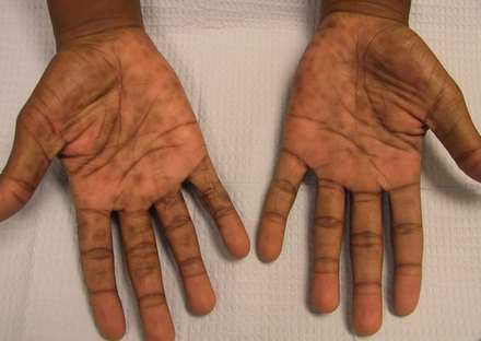
Prurigo Nodularis (hands)

Picture 2 – Prurigo Nodularis on hands

Prurigo Nodularis images

Image Source -askdrshah.com

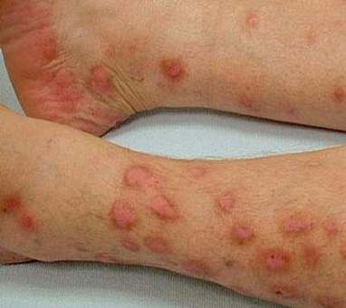
Prurigo Nodularis on legs

Picture 4 – Prurigo Nodularis on legs

image source – mf.cz

No responses yet

Next »

© 2017 eHealthWall.com. All Rights Reserved. Privacy Policy
This website is for informational purposes only and Is not a substitute for medical advice, diagnosis or treatment.