Archive for January, 2012

Penile Fracture (Broken Penis) – Symptoms, Causes, Treatment, Surgery, Pictures

Jan 13 2012 Published by under Diseases & Conditions

What is Penile Fracture?

Broken Penis is a fracture of the penis where it affects directly the Tunica albuginea. It is a tissue that envelopes and protects the corpora cavernous penis. The tissue Tunica albuginea is mainly involved in the erection of the penis with the help of the Buck’s fascia where deep dorsal vein restrict the blood from leaving the penis and then maintain the erection. Once the penis is traumatized, the tunica albuginea may be ripped off and cause a bleeding or hematoma on the area. This damage may be the reason why the penis will not erect because the tunica albuginea is broken.

Broken Penis Symptoms & Signs

  1. Rolling Sign – a firm, stiff, and palpable hematoma – the blood seeped out of the cylinder where it is enveloped by the tunica albuginea
  2. Pain in the penis
  3. Ecchymosis of the penis
  4. Audible popping sound
  5. Eggplant Deformity – the Broken Penis has the appearance of an eggplant where it is swelled, curved like the letter S, bruised, and angled off towards the other side of the penis.
  6. Extension of the swelling and hematoma to the scrotum, perineum, and suprapubic region
  7. Potential injury of the urethra – Dysuria, hematuria, urinary retention, blood present at the penile meatus

Broken Penis Causes & Riskfactors

  1. When a penis is accidentally traumatized or bended laterally more than it has to during an erection may cause pulling of the stiff tissue of tunica albuginea, therefore lacerating the area. In some instances, Broken Penis happens during a sexual intercourse. Intense and aggressive positions and acrobatic activities can give rise to the pulling and damaging of the tissue of the erected penis and flexing it beyond its strength.
  2. Aggressive masturbation may put the penis into damaging the tunica albuginea
  3. Taqaandan – a practice that is performed to detect the existence of erection by bending the head part of the penis while the shaft is erected. There will be a click that is felt and heard. It is similar to the cracking of the knuckles and supposed to be painless. This practice has been reported to cause penile fracture or broken penis.
  4. Gunshot at the genital area – approximately 35% of the genital injuries are caused by gunshot.
  5. Stab wounds at the genital area
  6. Penile Amputation – it involves the mutilation of own genitals. It is mostly associated with people who have psychotic disorders.
  7. Genital Skin Loss – an infection where in involves gangrene of the soft tissues of the genitalia. It is a rare occurrence but Fournier gangrene makes up the 75% of the cases of genital skin loss.
  8. Human or Animal Bites on the genital area
  9. Burns on the genital area

Broken Penis Diagnosis

The Broken penis is usually first seen and diagnosed by taking the health history of the patient, performing the physical examinations. Assessment of the Broken Penis by the health care provider should find the clinical manifestations before diagnosing Broken Penis. In some cases, to detect if there’s a presence of urethral injury in the Broken Penis, imaging studies are done such as

  1. Cavernosography – a routine specific radiographic visualization of the corpus cavernosum of the Broken Penis.
  2. Magnetic Resonance Imagery (MRI)
  3. Urethrographic Studies – this is to confirm or rule out the damage of the urethra and the extension of the trauma.

Broken Penis Treatment

a. Non-invasive medical management

1. Stabilization and Adequate fluids – to replace the blood loss to hematomas

2. The Broken Penis should be given proper compression, penis splints and bandages to avoid unnecessary painful erection of the Broken Penis and further damage the corpus cavernosum.

3. Cold compress – this is to relieve the Broken Penis from pain and hematoma

4. Administration of prescribed medication for the Broken Penis

  • Antibiotics are given to avoid the development of infection on the Broken penis
  • Estrogens that inhibits the erection the broken penis
  • Pain killers are also given to relieve the patient from the pain
  • Anticoagulants and thrombolytic drugs to avoid formation to much blood clot that may develop but should be use with proper caution to avoid uncontrolled bleeding.

5. Proper and Sterile Wound Care by cleaning the broken penis of dirt, debris, dead cells and wrapping with sterile gauze that is done every day (or as ordered by the doctor).

b. Invasive Surgical Management

No matter how small or big the trauma has inflicted the penis, surgical restoration of the penis is done to correct the damage done to the tunica albuginea, avoid the complications, erectile dysfunction, and urethral damage. Records show that 92% of the surgeries done had great results. There are three various types of incisions that are done to restore the broken penis:

1. Incision of the affected area to visualize the extension of the defect. It also provides examination of the nerve bundles that are affected in the broken penis.

2. Inguinal – scrotal incision is giving a great view of the broken penis’ root, base, and rear portion.

3. Circumferential – degloving incision is the one that is usually suggested by health care providers because it gives excellent cosmetic outcomes. This is the incision that provides the surgeon the entire exposure of the bilateral corpora cavernosa.

In some cases like penetrating trauma or presence of gangrene in the broken penis, the surgery should include these management:

  • The necrotic and gangrene tissue should be incised and debrided.
  • Intensive sterile cleaning of the area with antibiotic solution, creams, and Povidine.
  • Skin graft is done if the affected area is large and needed to be covered.

Prevention

Erection of the penis is inevitable because it is the penis’ nature to erect when stimulated sexually. One solution is to avoid the position that may cause too much bending of the penis. Also, broken penis occurs when it is banged against the partner’s pubic bone or any hard surfaces.

Pictures
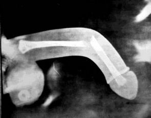
Broken penis picture (diagramatic)

Picture 1 – Diagramatic representation of broken penis (Truely, bone is absent in penis)
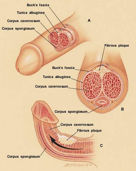
Penis anatomy structure parts picture

Picture 2 – Real anatomy inside penis

Image Source – peyronies-disease-help.com
No responses yet

Ice Pick Headaches – Treatment, Causes, Symptoms, Remedies

Jan 12 2012 Published by under Diseases & Conditions

What is Ice Pick Headache?

Ice pick headaches are classified under primary stabbing headaches cluster. Ice pick headache is named as such because it occurs like a series of stabs. It is also being referred to as “jabs” and “jolts”. Some medical professionals would refer it as opthalmodynia and periodica. Patient attacked by this kind of headache is unable to act or respond during the episodic attack. It is often misdiagnosed as migraine or cluster headaches for they have similar kinds of symptoms manifested by the patient. It is often seen in either male or female adults and rarely in adolescents or young children. It becomes more prevalent when one is in the middle age state. It might be due to the fact that they are more prone to the causes that will be mentioned as we go along this disease process.

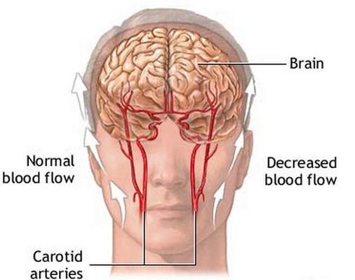
Ice Pick Headaches pathophysiology, brain image

Picture of Ice pick headache pathophysiology

Image source – Adam Inc

Symptoms

The symptoms or signs that are associated with Ice Pick Headaches are as follows:

  1. Painful headache
  2. Occurs in the head
  3. Sudden type
  4. Brief (within 5 to 30 seconds)
  5. Episodic headaches
  6. Piercing kind of pain
  7. On and Off pain
  8. No other disease condition associated
  9. No other symptoms

It is described as sudden, brief which usually last 5 to 30 seconds, episodic, short lived and recurring headaches, which are usually occurring at regular intervals at irregular frequency. It is also described as a piercing like pain. Patients would often report that they felt as if ice picks have been stabbed in their heads. It occurs mostly in the eye and orbit, which can be felt behind or around the eyes, temple, or a rare occurrence in parietal regions, which trigger the first division of the trigeminal nerve. It feels like you are being stabbed by ice picks, stabbing your head without warning, tending to go in and out of the blue. Person feeling such will report pain that’s sharp and severe leaving you incapacitated. Yet they don’t last long. There should be no accompanying symptoms and no underlying disorders detected.

Causes

There is no exact known etiology of why such headaches occur. Some says it is a neurologic idiopathic in nature. However some of these headaches are due to serious problems and some of them are not really leading to serious conditions. Yet recent studies found out that people experiencing migraines, also end up experiencing this kind of headache. Although they are occur independently from migraines. Another study suggests that inflammation, as another cause of this condition. It is said that it may cause adjustment to sensory nerves. Other triggers that may trigger migraine may also trigger ice pick headaches. In addition to that, researchers suggest that certain food, pollutants, and/or stress may lead to having this kind of disease.

Diagnosis

To be able to diagnose this condition, the symptoms mentioned above must all be met. First, you have to disregard the possibility of diagnosing it as a migraine or a cluster headache. The doctor usually diagnoses this kind of headache. You should consult the doctor when you manifest the symptoms early so that treatment will be given as soon as possible.

Treatment

One problem when you are dealing with this kind of headache is that they usually come and go. Treating it is difficult. One difficulty when you are to treat such headache is when the medication has taken its effect, the headache is already gone. This type of headache is felt either in day or at night. It is troublesome when one experiences it at night. It will disturb your sleeping hours.

Non Steroidal Anti Inflammatory Drug – Indomethacin

One drug that is often prescribed in this disease is Indomethacin, which is classified under NSAIDS or Non steroidal Anti Inflammatory Drug. However, Indomethacin causes discomfort in your digestive system. NSAIDS or non steroidal anti inflammatory drugs can aid in moderating the attacks. It blocks the so called COX enzymes and reduces prostaglandin. Thus, inflammatory process (calor, rubor, dolor, funtio laesa, and tumor) is kept to a minimum.

Anti Depressants

Another drug, which can be prescribed, may include anti depressants. Medication classified under this drug will improve your sleep. This is often prescribed to patients who have troubled sleeping at night due to the pain caused by this kind of headache.

Anti epileptic

Some doctors will prescribe an anti epileptic medication. It is also used to induce sleep. Helping you in achieving an uninterrupted and peaceful sleep.

Melatonin Hormone

The hormone melatonin, will aid people who are experiencing this headache at night. It will aid in having adequate peaceful sleep without interruptions.

Remedies

With regards to remedies that may help aid in minimizing or controlling the symptoms of the headaches, you can both try independent therapy or dependent therapy.
Independent Treatment

Relaxation Techniques

Under the independent therapy, you can try relaxation techniques. It is an independent technique. This can be done through remaining calm and through taking slow, deep breaths. Although in some cases, this kind of headaches will resolve on their own. You just need to discover, identify and remove the stressor that may cause recurrence of this disease condition.

Meditation

You can also try meditating. You can go to a silent and serene place to meditate. This will relieve you of stress.

Taking Good Care of Your Overall Health

The main important remedy to prevent recurrence or occurrence of this disease is to take care of your overall health.

Eat healthy

You try to eat healthy. It aids in the improvement of your immune system. Include fruits and vegetables. Eat salads. It will help in detoxifying your body.

Do Exercise

You have a proper exercise at least three times a week or probably through walking through stairs instead of using elevators.

Change your lifestyle

Change your lifestyle by staying away from stress as much as you can possibly can. You also try to get at least eight hours in day of uninterrupted sleep. This kind of activity will help in decreasing symptoms felt.

No responses yet

Torn Ligament in Knee, Shoulder, Ankle, Wrist – Symptoms, Diagnosis, Treatment

Jan 11 2012 Published by under Diseases & Conditions

What is a Ligament?

Before we can understand what a torn ligament is, we should first define a ligament. Ligament, by definition, refers to a group of tough, fibrous, stretchy and dense connective tissue  including attenuated collagenous fiber. Ligament elasticity is arranged in a crossing pattern that prevents the joint from being loose.

Ligaments are what connects one bone to the other bone in the body forming a joint. These ligaments join the bones and allows a person to walk without falling or tumbling over. It is different from what a tendon does, it connects bones to muscles. Ligaments limit the movement of a certain joint and sometimes prevents mobility altogether.

The ligaments in the joints do the strengthening in the articular capsule and stabilizes the joint. It also controls a range of and prevents dislocation and other causes of injury. Ligaments  have the characteristic of being viscoelastic, it means that a ligament slowly lengthen during tension and returns to their original structure when the tension is gone. However, returning to their original structure becomes impossible if it is stretched more than a specific point for long extended period of time.

What is a Torn Ligament?

Tears or strains in a ligament occurs if a joint becomes overstretched or is twisted. The most commonly affected area by tears or strains are ankles and knees. To prevent it, individuals, especially athletes, dancers, martial artists, gymnasts or even normal people must perform stretches and other exercises to lengthen and strengthen their ligaments making it more elastic and flexible.

Torn Ligament Symptoms

The symptoms for any kind of ligament injury will be similar, regardless on the extent of damage. Usually, there is a snapping, popping or a cracking sound during the injury, when there is a torn ligament.  The usual symptoms include swelling, bruising accompanied with severe pain and pressure on the joint. There is also instability—a feeling that the knee is locking or is about to break down. Movement is limited or totally absent in cases of a torn ligament. Standing is also difficult for those with a torn ligament, in which cases it can’t bear a full weight. Also, physical examination reveals that there is a dent on areas where the torn ligament is present

Torn Ligament at Various Places

Torn Ligament in Knee

Mainly anterior and posterior cruciate ligament injury cause torn ligament at knee. The most common cause for the anterior part is a stretch or tear or both by a sudden and twisting motion while the posterior injury is most often caused by a direct impact on the site such as a football tackle or motor vehicle accidents.

Torn Ligament in Shoulder

Glenoid labrum tear cause Torn Ligament in shoulder. The injury to the tissue rim that surrounds the shoulder socket occurs from a repeated shoulder motion or from an acute trauma. Athletes that throw  or weightlifter often experience gleniod labium tears due to repetitive motion of the shoulders.

Examples of injuries that could lead to trauma include:

  1. A direct shoulder blow
  2. A sudden pull when initiating on lifting a heavy object
  3. A violent reach over the head when trying to prevent a fall or slide
  4. Falling over on an extended arm

Torn Ligament in Ankle

Torn ligament in ankle caused by an unexpected joint twist. It can be a tear, stretch, or a complete rupture of the ligaments that join the bones of the ankle.

Torn Ligament in Wrist

Wrist injuries are becoming more common and becomes an annoyance given on how reliant we are on our wrists and hands. During a fall, the reflex is that individuals tend to use their hands to support or break a fall. This makes the wrist twisted or torque and this is commonly cauled as a wrist sprain.

Diagnosing a Torn Ligament

Thorough patient history will be taken, consisting of

  • Asking symptoms and what had happened, described in full detail.
  • Physical examination is done to examine the signs of a torn ligament, this also includes performing movements such as squatting, hopping or simply stepping. The examiner may also test for injury by flexing and bending the injured area. Also, the examiner feels for fluid in the joints by gently pressing over the kneecap (for knee injuries) if there isn’t much evidence of swelling.
  • Other tests may also be done to confirm the diagnosis of torn ligament. It includes MRI, ultrasound or X-ray. These tests help detect complicated or more severe injuries.

Torn Ligament Treatment

Treatment of torn ligament that will be received is dependent on the extent and kind of damage attained.

Immediate Care

Follow the RICE method; Rest, Ice, Compression, Elevate. Rest the injured area for the first couple of days, for about one to three days then gradually introduce movement as able. Remember not to push yourself too hard, but don’t immobilize it completely that it loses its tone. Ice application is an important aspect of the care. It reduces the swelling and bruising. However, do not directly apply ice to the skin because it causes real damage. In ice application, cold compress is used, it can either be an ice bag or frozen packs wrapped in a towel. To put pressure on the injured area, compress the joint by putting bandage to support it. It can also help decrease the swelling. Lastly, elevate the affected area above the level of the heart and keep it well supported.

During the first three days, DO NOT DO HARM (Heat, Alcohol, Running, Massage).  These methods add much damage to the injury. Heat involves using hot pack or even taking a hot bath.  Remember not to drink alcohol because it can increase bleeding tendency and swelling in the injured area. Do not run or do any form of exercises, pushing yourself too hard does not help in the recovery. Massage doesn’t always do well. In a torn ligament, massaging the injured area can cause more bleeding or swelling. Crutches or braces may be used to immobilize the affected area.

Medicine

Painkillers, usually Paracetamol or Ibuprofen can be used to treat mild to moderate pain. Although doctors may prescribe a stronger form of painkillers if necessary, in cases of severe pain. Anti-inflammatory and in combination with analgesics are more common to relieve swelling and inflammation accompanied by the pain.

Physiotherapy

During more severe or complex injury, individuals may be referred to a physiotherapist—health care professional that specializes in the movement and mobility.  In addition, even without referral, an individual can also choose to visit a private physiotherapist to develop an individualized program especially for the injured area. Rehabilitation exercises gradually strengthens the injures area as well as stretching the muscles for better improvement. Different kinds of techniques are used to speed up the healing process. Braces can also be used occasionally during rehabilitation if the injury is severe.

Surgery for Torn Ligament

In some cases, surgery of torn ligament is needed to repair the injury. This is likely if:

  • the lateral collateral ligament is ruptured
  • the anterior cruciate ligament is damaged and an individual is athletic, or if the medial collateral ligament is damaged or a cartilage is torn—reconstruction operation may be needed. This involves taking a tendon graft to replace the ligament that was damaged
  • the damaged ligament or tissue is more than one
  • the patellar tendon has been torn

Natural Treatment for Torn Ligament

Natural treatments for damaged ligaments are also possible. These involve herbs as a great alternative, herbs are safe and gentle on the body. Herbs are also beneficial for the overall well-being of an individual. Glucosamine sulphate  and Harpagophytum procumbens (Devil’s Claw) contains powerful  components that help promote health in joint and cartilage. Furthermore, Boswellia serata promotes muscle and joint comfort.

Torn Ligament Prevention

There are certain safety measures that can be done to reduce the risk of damage to the ligaments:

Regular exercise

Regular exercise to maintain a certain fitness level. The muscles in the body will be generally stronger and will be able to support weight in the joints. So it is good to be active and exercise and it is better if it is in a gradually increasing intensity.

If  an individual will engage in sports or rigorous exercise, a warm up exercise is advisable. It increases blood flow to the muscles and reduces the chances of taking damage or injury. Cool down is also necessary after the sport or rigorous exercise. However, the benefit of stretching, before or after exercise is still to be proved.

Torn Ligament Pictures

Torn ligament Knee pictures, pics

Picture 1-  Torn Ligament Anatomy

torn ligamnet ankle pictures

Picture 2 – Swelling of torn ligament at ankle

Torn ligament Knee pictures

Picture 3 – support for torn ligament

No responses yet

Greenstick Fracture Treatment, Symptoms, Causes, Diagnosis, Pictures

Jan 10 2012 Published by under Diseases & Conditions

What is a Greenstick Fracture?

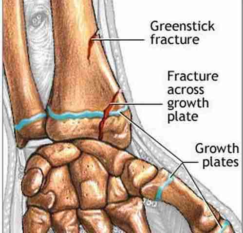
Greenstick Fracture is defined as a type of fracture where the bone bends and partially breaks. Greenstick fracture usually occurs during infancy and childhood when bones are still soft. This fracture was discovered by John Insall, a British-American orthopedist and Michal Slupecki, a Polish-American orthopedist who described the fracture like that of a green wood, which simply breaks on the outside when bent.

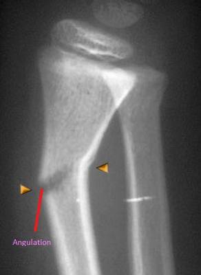
The bending is known as an angulation. The swelling caused by angulation might be dorsal, which signifies it can be found at the rear of the forearm. It might also be volar, which signifies that the swelling is found at the frontside of the forearm. Commonly, there will be rotation together with the bending force, which signifies that the bone will also be rotated along the bone’s axis and not simply bend a little.

There are three basic forms of greenstick fracture, namely:

  • transverse fracture
  • torus or buckling fracture and
  • bow fracture.

Transverse fracture occurs around the cortex that extends to the middle of the bone while torus or buckling fracture is caused by impaction, and the bow fracture occurs when the bone is curved along its longitudinal axis.

Greenstick Fracture at Wrist

These fractures are common at distal radius or distal ulna. It is like to happen in children who fell on a widely spread arm. Reduction will not be done if the dorsal angulation is mild and the reconstruction will start as the kid grows. A gradual and continuous pressure to lessen the deformity is known as reduction. It is performed until the intact dorsal cortex is fractured. If the cortex fail to break it may lead in intensifying deformity throughout the duration of the cast. The forearm is slightly turned around dorsally and a long arm cast is used for four to six weeks.

Greenstick Fracture at Radius

The bones of the forearm are radius and ulna. The fractures may occur in three sites: proximal, medial or distal end of the bone. Greenstick fracture in this area is usually shown by an incomplete break with arm displacement.  Greater force may cause complete break and therefore must be given proper medical attention.

Greenstick fracture of radius (wrist)

Picture of greenstick fracture of radius at wrist region

Greenstick Fracture Causes

Children are always active, they love activities such as running, playing, tumbling and skipping.  Thus they always sustain injuries and most likely to experience fracture. These are the events when fractures may occur:

  • Playing contact games like hide and seek
  • Playing in high places
  • Tripping
  • Running on uneven surfaces
  • Putting much weight on the limb

Usually the harmed part is the arm bones because of the tendency to guard or throw out the arms to stop oneself from falling. Such force causes the arm bone to bend and to break, but not completely. Many kids experience breaking a limb at some time in their lives and greenstick fracture is usually nothing to be concerned of, but it requires medical attention.

Greenstick Fracture Symptoms

There will be no symptoms in some cases but there would be always symptoms emerge like:

  • Intense pain
  • Swelling of the affected area
  • Presence of abnormally bent or twisted limb
  • Slight bruising on the affected area
  • The child guards the affected part
  • Decreased range of motion on the injured part

Diagnosis

The main factor about greenstick fractures is that it can go unnoticed and undiagnosed. A thorough medical history and physical exam is performed to confirm the diagnosis. Diagnostic tests like X-ray to reveal greenstick fractures are done to the injured and uninjured limb for comparison. In some cases were greenstick fractures are difficult to view, tests like ultrasound or computerized tomography can also be done to provide better visualization.

Greenstick Fracture X-ray picture

X-ray showing angulation/bending in Greenstick fracture

Treatment

Greenstick fractures treatment requires fracture reducing. This is done by:

  • Slightly pulling the bone apart and putting it into place to straighten it. To make sure that the fracture will heal correctly, it is needed to be immobilized.
  • Usually, casts are used or a removable splint for the convenience of the child. Since children’s bones tend to heal faster than those of adults, the cast or the splint maybe removed in a short span of three to four weeks.
  • Physical therapy is also indicated to regain mobility and movement of the affected limb. Mostly, this type of fracture heals completely after being reduced and placed in a cast. However, if there are some instances that these measures do not take effect, surgery is advised to correct the fracture.
  • When surgery is done, post operative traction may be used to straighten the hard fracture

This type of fracture is not as painful as the others, especially once the fracture has been reduced. In order to reduce swelling on the affected site, anti-inflammatory drugs can be given. Younger children may not able to express their feelings in the event greenstick fracture occurs or they can cry uncontrollably with the intensity of pain. Often they usually guard and become protective over the affected area. That is why parents are advised to have their child seek consultation when they suspect that their child sustained a major fall.

Prevention

A way to prevent greenstick fractures is

  • Supervision when your child is playing and keeping him or her from harm.
  • Use of car seats and seat belts are also advised.
  • Ensuring of wearing a safety gear for the children when playing is also important to reduce the risk of fractures.
  • The children’s diet must include adequate amount of calcium to keep bones strong and healthy.

No responses yet

Broken Ankle – Pictures, Symptoms, Treatment, Recovery, Rehabilitation

Jan 02 2012 Published by under Diseases & Conditions

What is Broken Ankle?

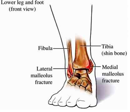
Broken ankle is defined as any fracture that happens in the distal end of the tibia and the fibula, and the talus bone of the foot. These three bones form the ankle joint and the talus is the only bone that is a part of the ankle joint. Tibia and fibula are parts of the lower leg.

Broken ankle is a very common injury and the severity of the fracture varies. Ankle fracture may be partial, or incomplete, open or closed which may be a result for fall, collision/impact, or accident. Thorough assessment and an x-ray examination and/or CT scan, is done by an orthopedic doctor to review the injury and the extent of damage to the nerves and surrounding tissues.

Broken Ankle Symptoms

These common signs and symptoms for broken ankle are

  • Pain to touch with limited movement of the ankle
  • Swelling and/or bruising on the affected area
  • Inability to walk or stand and bear weight on the ankle
  • Numbness or tingling sensation on the affected ankle
  • Deformity around the ankle
  • Broken skin with visible bone particle

Broken Ankle or Sprained Ankle?

In a broken ankle the bone is fractured. In a sprained ankle, the ligaments are over-stretched or torn. The signs and symptoms are often similar. Here are some hints in distinguishing the difference between the two.

  • What caused the injury? If your ankle gets a twist or if you lost your balance as you stepped on an uneven surface, it would probably be a sprain. Heavy impact causes bone break.
  • If your ankle swells but you are still able to stretch it, that’s an ankle sprain.
  • If the pain is severe, accompanied by swelling, bruising and tenderness, that’s a broken ankle.
  • Numbness is indicative of broken bone.
  • Stand up, take a few steps. If you are able to walk, it indicates that you have a sprained ankle.
  • Look at the affected area. If the joint appears kinked, or uneven, the bone must have been broken.
  • Try to make the injured person move the joint. A broken ankle cannot be moved, a sprained ankle can be moved even if it is painful.
  • Seek professional advice to be certain of the injury acquired.

Broken Ankle Treatment

Anyone with a broken ankle must be checked by an orthopaedic doctor for the treatment plan to be established.

  • The first aim is to minimize the swelling to control the pain and minimize damage to surrounding tissues. After the x-ray, the doctor can tell how bad the fracture is.
  • Splinting of the ankle is done initially to immobilize the fracture and it will be kept in place for a few days to give room if the swelling persists. The ankle will be aligned properly prior to splinting.
  • If the swelling subsides, casting will be done. Casts are made either of plaster or fiberglass.  Plaster cast molds better with the skin and is widely use to hold the bone in place. However, if the fracture is unstable and some healing has already taken place, a fiberglass cast may be used. It is lighter and tougher to use. The whole body will not be immobilized, just the fractured ankle.
  • Crutches are used to assist in weight bearing as the ankle will not be used for weeks or months depending on the fracture. For the severe cases of broken ankle, surgery is required. The maintenance of the proper alignment of the fractured ankle is of utmost importance for the ankle to heal and be utilized as it was before.

Broken Ankle Surgery

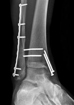
A general anaesthesia will be given to the patient, and then a cut on the skin near the ankle will be done. Special screws and plates will be used to put the bones together and hold it in place while the bone heals.

  • Then, the skin will be fixed with the use of stitches. A temporary plaster cast will be molded just below the knee up to the toes to secure the ankle in proper alignment and keep it immobile.
  • The wound may be painful and the doctor may give painkillers to ease the pain as well as antibiotics to prevent infection. The surgeon who performed the operation will give instruction as to when the ankle may be moved.
  • Some would prefer to get the fractured ankle moving before sending the patient home in around 2-3 days. Appointment will be scheduled. In 10 days, the stitches will be removed and the ankle will be in plaster cast for approximately six weeks. For the first two weeks, the affected leg should be elevated at rest several times a day to minimize the risk of further swelling as the cast may impede with the circulation once the ankle swell.
  • Crutches may be used to move around with support, avoiding weight on the fractured ankle. Once at home, utmost care for the fractured ankle should be done. Plaster cast should not get wet, cut, or pressed on. Nothing should be put underneath the cast and the skin inside should not be scratched by anything.
  • If the toes get blue, swollen, and cannot be moved, or at the presence of numbness or severe pain, go back to the hospital immediately. The screws and plates may or may not be removed but if it becomes uncomfortable, it may be removed 18 months after the operation. If the plaster cracks, or becomes soft and loose, it should be brought back to the plaster technician.

Broken Ankle Recovery

If the broken ankle is clean, no shifting of the bone and doesn’t require surgery, the broken ankle will heal within a six week period with proper medical management. A repair with the use of invasive surgery would have the broken ankle heal in about eight to 10 weeks. For this long period of recovery, you will need a

  • lot of patience
  • entertainment
  • emotional support
  • lots of pillows
  • easy access to food and drinks
  • lots of strength
  • pain medication and
  • bath chair
  • Do not attempt to walk unless your doctor told you so.

Rehabilitation (Physical Therapy)

Rehabilitation is very important. When your physician said that you may start moving your ankle, physical therapy and home exercises are available and you should start doing it regularly. Eventually, you will also start doing exercises to strengthen the surrounding muscles of the ankle. It may take several months for the muscles around your ankle to get strong as before the injury took place. Even after the fracture has healed,an ankle brace may still be worn for several months. Both of your ankle support almost all of your body weight, so it is very important getting them back to full strength again. Here are some of the therapeutic exercises for the recovering ankle:

Ankle Rotations

This exercise helps move the ankle joint, makes good circulation and loosen ligaments that have been inactive while on cast. Sit on a chair, hold your leg just above your injured ankle crossed over your healthy leg. Slowly rotate your foot at the ankle, turning it in circles. Do only as much rotation and stretching you feel comfortable with. If you feel pain, stop and rest.

Towel Scoop

To help achieve range of motion in your foot, sit on a chair barefooted and a hand towel on the floor in front of you. Sit on a surface and use your injured foot to scoop the towel from side to side, keeping your leg still. Do it several times a day.

Wall Stretch

Wall stretches can help the ankle and the muscles associated with it. Stand several feet away from a wall, pressing your hands to it. As if you are trying to push and walk through the wall, with the injured foot on the floor and knee straight behind the healthy foot with knee bent. You should feel a stretch. Hold stretch for 10 to 15 seconds. Stop if you feel pain.

Broken Ankle Pictures

Picture of Broken ankle anatomy Picture 1 – Pic of broken ankle anatomy
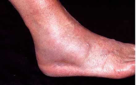
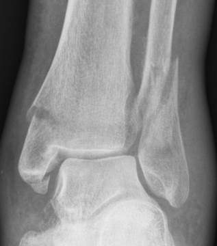
Image source – empowher.com

picture of Broken ankle showing swelling, redness

 Picture 2 – Broken ankle showing swelling, redness

Image source – sciencephoto.com

X-ray of broken ankle

Picture 3 – X-ray showing broken ankle (tibia, fibula)

Image source – footankle.com

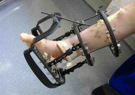
External fixation of broken ankle

Picture 4 -External fixation of broken ankle (x-ray view)

Ring fixator for Broken ankle

Picture 5 – Ring fixator for Broken ankle

No responses yet

« Prev

© 2017 eHealthWall.com. All Rights Reserved. Privacy Policy
This website is for informational purposes only and Is not a substitute for medical advice, diagnosis or treatment.