Archive for February, 2012

Sleeping Sickness

Feb 06 2012 Published by under Diseases & Conditions

What is Sleeping Sickness?

Do you always travel in Africa? If yes, are you aware about the Sleeping Sickness or so called by the medical experts as African Trypanosomiasis or Congo Trypanosiomiasis. This is a disorder caused by small parasites that are transmitted to human hosts by bites of infected tsetse flies (Glossina Genus). The disease develops slowly and if treatments are delayed, it can lead to a serious infection in the brain that can lead to fatality or death. In addition, it can be transmitted through mother to child, blood transfusion and sometimes infections through handling blood of an infected person.
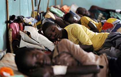
sleeping sickness pictures

Sleeping Sickness Causes

What causes the person to be infected?

  1. It is caused by infected tsetse flies that carry 2 types of germs: Trypanosoma brucei rhodesiense (East African sleeping sickness) and Trypanosomoa brucei gambiense (West African sleeping sickness).
  2. Sleeping Sickness normally is present in rural areas, living in woodlands, rivers, streams and forest.
  3. Both male and female tsetse flies can transmit the infection and they bite during daylight hours.
  4. Once you are bitten, the infection will spread through your blood and if it is not detected early it can affect the central nervous system which is fatal to the person who is infected by the disease.

Sleeping Sickness Symptoms

First Stage (Haemolymphatic phase)

In first stage, Haemolymphatic system is affected. The symptoms are

  1. Fever
  2. Indurate chancre at bite site
  3. Headache
  4. Joint pains
  5. Itching
  6. Winter bottom’s sign is clearly visible: swelling of lymph nodes (lymphadenopathy) along the back of the neck, which indicates inflammation.

Second Stage (Neurologic phase)

In second stage, Central Nervous System is affected. The symptoms are

  1. Confusion
  2. Loss of appetite
  3. Reduced coordination
  4. Disruption of the sleep cycle (daytime somnolence followed by night time insomnia)
  5. Irritability
  6. Tremors
  7. Muscle rigidity and tonicity
  8. Stupor and Coma (which gives the name “sleeping sickness”)

Sleeping Sickness Diagnosis

The best way to diagnose if the patient is positive for having a sleeping sickness disorder is through identification of parasites in the patient sample through microscopic examination. Patient sample that can be used in diagnosis include blood smear, cerebrospinal fluid test, lymph node aspiration, and bone marrow aspiration.
The second way in diagnosing a person is through physical examination.

Sleeping Sickness Treatment

First phase

Pentamidine and Suramin these are drugs that are traditionally used in patients with first stage of sleeping sickness disorder.

  1. Pentamidine is an antimicrobial medication that is administered through injection and inhalation. It has many side effects like decrease in blood pressure, irregular heartbeats, heart failure, liver inflammation and some neurologic effects such as dizziness, confusion and hallucination.
  2. Suramin is an inject-able medication that causes parasites to lose their energy and die. The common side effects are diarrhea, headache, nausea and vomiting.

Second phase

Eflorinithine and Melarsoprol are drugs used in patients with second stage of sleeping sickness disorder

  1. Eflornithine is a new drug for the sleeping sickness disorder known for having a fewer side effect, more effective but costly. It is a drug that is ideal for killing susceptible bacteria responsible for sleeping sickness.
  2. Melarsoprol is a highly toxic drug that is administered through injection and it is supervised by a physician as it produce a similar effects as arsenic poisoning.

Sleeping Sickness Prognosis

Untreated infection both the Trypanosoma brucei rhodesiense (East African sleeping sickness) and Trypanosomoa brucei gambiense (West African sleeping sickness) is fatal that it might lead to death. So, therefore both of them must be treated without delay. The blood borne stage or the haemolymphatic stage of infection is the opportune time to treat the disease. If the disease is left untreated, or the parasites already invades the central nervous system and the patient already show signs of neurologic symptoms or when the patient is already experiencing the sleeping sickness the disease is invariably fatal, with progressive mental deterioration leading to coma and death.

Sleeping Sickness Complications

  1. Cardiomyopathy: disease that affects the muscle of the heart
  2. Heart Failure: is a serious condition where the heart continues to beat but it’s too weak to pump sufficient amount of oxygen-rich blood to and from the lungs and to the rest of the body.
  3. Coma: is a deep state of unconsciousness which the patient fails to respond normally to painful stimuli, lights or sound, and does not initiate voluntary actions.
  4. Death

Sleeping Sickness Prevention

  1. If you came to a place where tsetse flies are present and you experience symptoms like fever, headache and some disturbance in your sleep pattern, you immediately seek a doctor for diagnosis and treatment. Diagnosis includes different types of test to confirm the presence of infection like blood smear or complete blood count and will conduct physical examination for the diagnosis of central nervous system disturbance. The treatment of this disease is a far-reaching process so it needs the attention and willingness of the person to be treated.
  2. Wear long sleeved shirt and pants in medium-weighted and in neutral colors. Tsetse flies are easily attracted to bright and dark colors.
  3. Inspect vehicles before entering because these flies are attracted to motion and dust.
  4. Avoid bushes. Tsetse flies don’t bite during the hottest time of the day but if they are disturbed they will bite.
  5. Using insecticides and clearing shrubbery helps in disturbing the breeding of the flies.
  6. Improving housing and sanitation. This can result in reduction in the rate of infection.

Remember

  1. There are still no vaccines or drug to prevent Sleeping Sickness.
  2. Sleeping sickness should be treated as early as it’s detected because if not, this disease will worsen as days and weeks will pass because the infection will spread in the body that will result to irreversible cure that can lead to death.
  3. Preventive measures are aimed by both government and health organization in minimizing contact with tsetse flies.

No responses yet

Ludwig’s Angina

Feb 04 2012 Published by under Diseases & Conditions

What is Ludwig’s Angina?

Ludwig’s angina is a life-threatening infection of the connective tissue of the submandibular or sublingual area (located on the floor of the mouth). It was named after Wilhelm Friedrich von Ludwig, a German physician who first knew about the disease in 1836. Ludwig’s angina is also known as angina ludovici, angina maligna or morbus strangularis because of the inflammation it causes in the floor of the mouth causing a strangulating feeling (from the word angina meaning “strangling”).

Ludwig’s angina is different from angina pectoris or chest pains. However, chest pains may be present in advanced cases of Ludwig’s angina, where the infection reaches the retrosternal space.

Ludwig’s angina is simply known as submandibular and sublingual space infection.

Ludwig’s Angina Pathophysiology

Ludwig’s angina starts from a dental infection. Almost half of the patients suffering from Ludwig’s angina have existing or previous infections of the teeth and gums. Periodontal infections are commonly caused by streptococcus and staphylococcus organisms which are present in the mouth even when the person is healthy.

Once the bacteria travel and spread, it reaches the submandibular and sublingual areas causing inflammation and swelling. Because of the inflammatory response, the body produces chemical mediators responsible for pain, erythema and swelling, which are evident to a patient. The infection and inflammation extends to adjacent tissues such as the pharynx and neck. Abscess formation develops usually on these areas. Patients typically breathe through the mouth as a result of swelling of the airways. Because of inflammation of the tongue, it falls back at the base which causes airway obstruction. Massive swelling also makes the upper airways constricted.

The infection further reaches the lungs and mediastinum. Retrosternal abscess develops and results in chest pains and chest heaviness. Descending infections in the lungs cause pneumonia and formation of pulmonary secretions that further aggravates the difficulty in breathing. More severe cases may eventually lead to sepsis and shock.

Ludwig’s Angina Symptoms and Signs

Symptoms of Ludwig’s angina include:

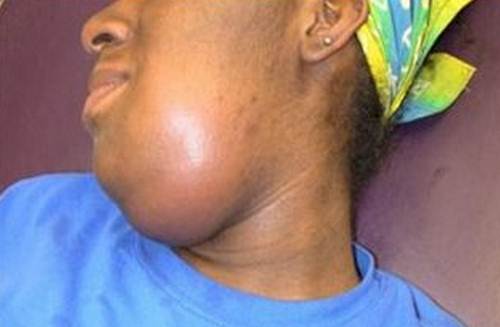
  1. Swelling of the submandibular and sublingual areas
  2. Pain on the tongue and neck
  3. Redness and swelling of the neck
  4. Fever
  5. Malaise, weakness and fatigue
  6. Dysphagia or difficulty in swallowing
  7. Drooling
  8. Difficulty breathing
  9. Tachypnea (rapid respirations)
  10. Stridor (a harsh sound heard during inspiration which signals an obstructed airway)
  11. Confusion or mental changes due to hypoxia
  12. Earache as a sign of spread of the disease to the ears
  13. Unusual speech

Contact a health provider when severe symptoms such as stridor, shortness of breath and chest pains occur.

Picture 1 – Observe swelling at submandibular region

Ludwig’s angina Causes & Risk Factors

Bacterial Causes

Ludwig’s angina is generally caused by a streptococcal bacterial infection, but other microorganisms can also cause it such as Staphylococcus, Bacteroides, Pseudomonas, Escherichia coli, Klebsiella, Enterococcus, Candida and Clostridium species. Since it is primarily caused by streptococcus bacteria, it is considered a form of cellulitis in the submandibular and sublingual areas.

Other Factors

  1. Submandibular swelling is most often associated with periodontal infections such as tooth abscess and is responsible for about 90% of the cases.
  2. Other risk factors include previous neck trauma, mandibular fracture, parapharyngeal infections, tongue piercing and even neoplasms, all of these may introduce microorganisms to the mouth and cause infection.
  3. People with immunocompromised status such as in HIV, organ transplant recipients, diabetes mellitus, alcoholism, glumerulonephritis, anemia, blood dyscrasias and systemic lupus erythematosus have higher risk in developing the disease.
  4. Adults and elderly are the most common age group who suffer from the diseases as this is uncommon to children.

Ludwig’s Angina Diagnosis

Radiology

Single plate x-rays are sometimes not efficient in determining the extent of the infection. A Computed Tomography (CT) scan of the neck and head is the best exam to determine areas affected such as the upper neck, chin and floor of the mouth. This may also assess spread to the thoracic area. Ct scans involve the use of several x-ray beams to check all angles of the area being examined.

Bacterial Culture

Fluid samples from the affected areas are taken to determine specific microorganisms that cause the infection. Almost half of the cases are caused by streptococcus bacteria.

Ludwig’s Angina Treatment

Ludwig’s angina is a life-threatening disease, but can be treated using appropriate therapy. Most of the mortalities in the earlier years are associated with poor diagnosis and lack of antibiotics to treat the disease. Today, advanced diagnostic procedures and antibiotics had made Ludwig’s angina as a rare disease. The primary treatment for Ludwig’s angina is the administration of antibiotics and airway management.

Antibiotics

Antibiotics are given to eradicate bacterial infection. Penicillin is the drug of choice, but other broad spectrum antibiotics can be used such as macrolides and cephalosporins. Proper allergy testing is done to avoid anaphylaxis since the antibiotics are usually given via the intravenous route. Broad spectrum antibiotics are given initially when the result of the culture and sensitivity is not yet available. These antibiotics are those who are able to kill several bacteria including gram negative and gram positive ones. When culture and sensitivity testing is available, then a more specific antibiotic is given, one in which the causative microorganism is most sensitive. Ludwig’s angina antibiotics are given for several days depending on the severity of the infection. It may be shifted to oral antibiotics as long as the patient is able to swallow and breathe effectively.

Steroid Therapy

Steroids have been used for its anti-inflammatory effects. Steroids significantly reduce inflammation on the site and improve the breathing pattern. However, steroids have not been used extensively today because it decreases the immune system that further leads to more infection.

Airway Obstruction Management

Airway management is essential in saving the life of the patient. Obstruction in the airways is a medical emergency and if airway obstruction is present, emergency measures should be carried out immediately. A tracheostomy tube or cricothyroidotomy can be placed by surgically creating an opening on the anterior neck into the trachea or wind pipe. A tube is then inserted to maintain the airway patent. Aside from this, an endotracheal tube may also be done in more severe cases of obstruction. This involves the insertion of a flexible tube into the mouth or nose going to the lungs. A guided intubation is used because blind intubations may lead to injury of the nasal, oral and tracheal mucosa. Secretions are suctioned as necessary to prevent accumulation and hypoxia. Characteristics of secretions are also assessed to determine infection. A greenish to yellowish phlegm indicates bacterial infection of the lungs. Breath sounds should be assessed every now and then to determine proper entry of oxygen.

Surgical Management

More than 50% of Ludwig’s angina cases develop swelling that is suppurative. Because of this, imaging studies are needed such as CT-scans to determine the extent and site of the complication. Most common site would be the neck. Drainage of the neck is done to remove accumulated fluids or pus. Incision and drainage is a minor operation that requires a sterile technique to prevent re-infection.

Supportive Management

Tooth extractions and dental treatment is also performed to stop the root cause.

Ludwig’s Angina Complications

Submandibular, sublingual swelling and inflammation leads to potentially life-threatening complications such as:

Airway Obstruction

This is the most common complication of Ludwig’s angina. As the mouth and the throat inflames, the airways become obstructed. Patients often exhibit mouth breathing and the tongue often falls back causing obstruction.

Sepsis

When the infection becomes severe, the microorganisms travel through the circulation and cause infection to other parts of the body. They grow and proliferate on the blood which causes bacteremia and finally lead to septic shock.

Mediastinal and Pharyngomaxillary Space Extension

Bacteria spread on to adjacent structures and spaces. When the mediastinum is affected, patients usually complain of shortness of breath and chest pains.

Osteomyelitis

Microorganims reach the bones through the circulation and significantly cause infection.

Ludwig’s Angina Prevention

The most effective prevention is to have regular dental check-ups to prevent periodontal diseases. Dentists should be consulted every six months for prophylactic therapy. When tooth and mouth infections occur, they should be treated accordingly with antibiotics to prevent spread to the rest of the oral cavity.

Ludwigs Angina Pictures

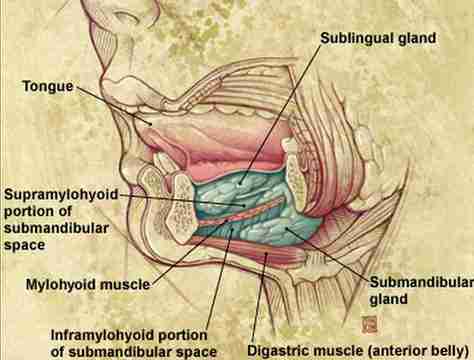
Anatomy of sublingual & submandibular region

Picture 2 – Anatomy of sublingual & submandibular region

Gross swelling at submandibular region in ludwigs angina

Pic 3 – Observe Gross swelling at submandibular region in ludwigs angina condition

No responses yet

Acrocyanosis – Causes, Symptoms, Treatment in Newborns and Infants

Feb 03 2012 Published by under Diseases & Conditions

What is Acrocyanosis?

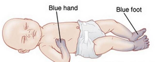
Acrocyanosis is the persistent bluish discoloration of the extremities including the hands, feet and parts of the face, but the most common is cyanosis of the hands. Acrocyanosis occurred some 100 years ago, but the nature of the condition is still unclear. Acrocyanosis has been thought to be similar with other conditions involving the cyanosis of extremities such as frostbite, reynaud’s disease, erythromelalgia, pernio and blue finger syndrome. However, acrocyanosis is not the same as to the pathophysiological mechanism. The extremities may also be persistently cold and cyanotic. Swelling and sweating of the extremities may also be present. Unlike other arterial occlusive disorders, acrocyanosis is not painful, ulcers and changes in the skin integrity also do not occur.

Acrocyanosis in Newborn

Acrocyanosis is commonly seen in newborns, whether preterm or full term. The phenomenon is considered normal to newborns because of immature circulation and underdeveloped capillaries. Acrocyanosis is one assessment in the early stages of newborn life especially during the first five minutes of life. Acrocyanosis may be present upon delivery of the newborn, but disappears after five minutes of spontaneous respirations. However, some institutions provide supplemental oxygen as precautionary measures for any underlying congenital problems.

Acrocyanosis Symptoms in Newborns & Infants

Observe Blue discolored hands and foot in acrocyanosis in newborn

Acrocyanosis in Infants

When babies grow, the circulation normally develops and infants do not usually present acrocyanosis. When acrocyanosis in infancy is still persistent, further evaluation should be done to check for congenital anomalies such as heart problems.

Acrocyanosis Pathophysiology

The exact etiology of acrocyanosis is unknown, but a benign neurohormonal condition may cause the disease. The condition results in narrowing of the arterioles on the proximal part of the extremities leading to cyanosis and cold, clammy skin.

When the arterioles go into vasospasms, the arteries and arterioles produce cyanosis. The persistent vasoconstriction in the area creates a local hypoxic atmosphere, which releases adenosine into the area. Because of vasospasm, adenosine enters the capillaries and causes vasodilation of the venules. The dilatation of the venules and constriction of the arterioles creates a countercurrent exchange system that causes heat to be retained. When this happens, profuse sweating of the palms and soles may occur as a result of excessive heat retention. Dilation of the veins also increases capillary permeability leading to extravasation of plasma into the interstitial space causing edema of the affected part.

Aside from adenosine, increase in serotonin also plays a role in acrocyanosis although the exact mechanism is not understood. Children taking Selective Serotonin Reuptake Inhibitors (SSRIs), a type of antidepressant, have manifested acrocyanosis as one of the side-effects. Other medications may also lead to acrocyanosis.

Acrocyanosis should not be seen as a serious condition, but precautionary measures and consultations should be taken as this may be caused by other underlying diseases.

Acrocyanosis Causes and Risk Factors

Exact mechanism of acrocyanosis is unknown. It is considered a benign neurohormonal disorder that causes narrowing of the arterioles at the upper and lower extremities.

Acrocyanosis is more common in children and young adults and the prevalence is also greater in women than in men. Studies suggest that risk factors to acrocyanosis include working outdoors, cold climate, and a low body mass index. Low body mass index explains why children, young adults and women have higher risk in developing it. The occurrence of acrocyanosis decreases with age regardless of the climate.

Women usually are relieved of the acrocyanosis after menopause, which implies that hormonal influence is a factor in its development.

Types of acrocyanosis

Acrocyanosis types depend on the causative factor.

1. Primary acrocyanosis

Primary acrocyanosis is the occurrence of the phenomenon without underlying cause or disease. These may be seen on newborn and young children with no apparent pathological condition that may lead to acrocyanosis.

2. Secondary Acrocyanosis

This type is usually caused by pathologic conditions such as arterial occlusive diseases in adults and heart anomalies in children and the newborn.

Acrocyanosis Symptoms and Signs

Acrocyanosis is seen as consistent bluish to purplish discoloration of the hands and extremities. Other signs and symptoms of acrocyanosis include:

  • Symmetrical discoloration of the hand and feet
  • Cold, clammy extremities
  • Swelling of the extremities
  • Sweating on the palms and soles which may be moderate to profuse

Despite those, people with acrocyanosis often have normal rate, amplitude and rhythm of peripheral pulses. Pain is also not present as compared to other occlusive diseases. The absence of pain and presence of normal pulses indicate that there is no significant problem on the circulation. Some individuals are asymptomatic, except from bluish to purplish discoloration of the hands and feet, which prompt them to seek consult and treatment knowing that it is a sign of a serious illness.

Acrocyamosis Diagnosis

Diagnosis of acrocyanosis involves extensive medical history and physical assessment. Other laboratory and imaging test is often not required. Presence of normal characteristics of pulses helps in ruling out the occurrence of arterial occlusive and peripheral artery diseases, such as Reynaud’s phenomenon. In occlusive diseases, the arteries are obstructed or narrowed which leads to impairment in the circulation.

  • Pulse oximetry also indicates a normal oxygen saturation of 95 to 100%. In people with peripheral arterial diseases, oxygen saturation results are commonly lower than 95% as a sign of hypoxia to the area.
  • Aside from the pulses and oxygen saturation, the skin is also observed for trophic changes. Acrocyanosis does not lead to skin changes and ulcers, aside from the cyanotic discoloration.
  • Some may require other tests such as capillaroscopy (visualization of the capillaries) to arrive in a definite diagnosis especially when connective tissue disorders are also present.

Acrocyanosis Treatment

Since acrocyanosis is not a disease, there is no definite treatment for it, although the following may help reduce the symptoms:

Prevention of Cold

The primary treatment is prevention of cold to avoid vasoconstriction and promote proper circulation.

Sympathectomy

Sympathectomy is a surgical procedure that destroys the nerves in an area that innervates the sympathetic nervous system. It is done in acrocyanosis to promote vasodialtion. However, it is rarely done because it is an invasive procedure and acrocyanosis really do not cause significant diseases.

Vasoactive Drugs

Calcium channel blockers and alpha adrenergic blockers (antihypertensive medications) become an option for the management, although it is not concurrently used. One study reveals that vasoactive drugs have little evidence that they are effective in the management of primary acrocyanosis which led to doctors not prescribing the drug.

The management of acrocyanosis simply focuses on the avoidance of cold. Health practitioners should also explain to patients that acrocyanosis without underlying arterial occlusive disease is not a medical emergency. It only needs supportive management.

Acrocyanosis Complications

Acrocyanosis often do not lead to complications and there is an excellent prognosis. The cause is benign which means no further developments of the disease is likely to occur. However, when acrocyanosis co-exists with other arterial occlusive diseases, then complications of the underlying conditions may arise such as skin ulcerations, infection and even amputation. Unless acrocyanosis occur from another condition, there is no possible risk for complications and disease. Patients can expect to have a normal life. Secondary acrocyanosis treatment also depends on managing the underlying cause.

Acrocyanosis Prevention

Although acrocyanosis has not been found to directly be caused by certain factors, prevention can still be implemented. Prevention for acrocyanosis includes the following:

  • Avoid cold temperatures – Cold temperature leads to constriction of the arterioles and possibly causes acrocyanosis. Stay inside during cold weather and wear appropriate clothing and gear such as gloves, boots, and coat.
  • Engage in exercise – Exercise promotes good circulation and oxygenation of cells. Concentrate on the upper and lower extremities.
  • Avoid constrictive clothing, stockings or shoes. – Promote proper circulation by ensuring that the extremities are relaxed. Use support stockings to enhance venous return and arterial blood flow.
  • Massage the hands and feet. – Massage relaxes the muscles and the underlying blood vessels thus preventing vasospasms
  • Avoid Stress – Stress stimulates the sympathetic nervous system which further leads to fight and flight response. Vasoconstriction occurs because of this. Learn some stress reduction activities such as guided imagery, focused breathing, gardening, interacting with other people and even avoidance of the stressor.

No responses yet

Hepatotoxicity

Feb 02 2012 Published by under Diseases & Conditions

What is Hepatotoxicity?

Hepatotoxicity, may sound very medical, but in layman’s term it is just liver toxicity. This term came from the greek word hepar which means liver and toxicon which means poison. It is the ability of a substance, typically hepatotoxic medications, chemical or drinking alcohol, to possess a damaging result or injury on the human’s liver.

The symptoms differ based on the extent of exposure and also the degree of the total damage or injury in the liver. If the damage is mild it may result to few symptoms while if the damage is severe it can eventually lead to failure of the patient’s liver.

Hepatotoxicity Causes & Riskfactors

Experts believed that the main reason for having Hepatotoxicity is the consequence of

  1. exposure of poisons
  2. harmful agents
  3. toxic substances and
  4. biological hazards, etc. at any time of the patient’s life.

It can also be brought about by

  1. previous liver problems
  2. abdominal diseases and digestive diseases
  3. adverse reactions of medications
  4. toxicity
  5. poisoning and
  6. overdose from medications.

I will now try to elaborate Drug Induced Hepatotoxicity and different drug interactions that can cause Hepatotoxicity.

Hepatotoxic Drugs

There are drugs, substances, medications, or toxins that can be toxic to your liver that eventually when you use religiously may lead to Hepatotoxicity. It is always advisable that you ask your doctor of any prescribed drugs and medications for your illness management can be hepatotoxic. This comprises of over-the-counter drugs, prescription medications, food supplements, herbal remedies or alternative medicines. There are few example of hepatotoxic drugs like Dacarbazine, DTIC-Dome, Sandimmune, Consupren and Sandimmun Neoral. But as I said a while ago it is very important to ask your doctor about every drug that you take.

Drug Interactions

Drug Interactions that can lead to Hepatotoxicity- It is not only hepatotoxic drugs that can cause hepatotoxicity, but some drugs when combined may react and in the end also lead to hepatotoxicity. These are only a few examples of drug combinations that can lead to liver toxicity:

a. INH and Tylenol

b. Isotamine and Tylenol

c. Laniazid and Tylenol

d. Nydrazid and Tylenol

This is why when you’re taking drugs or medications you have to be very careful. Be truthful to your doctor on what medications are you taking when he will prescribe your medication. Ask your doctor about drug interactions, it is better to ask than regret it in the future that you have not done any precaution.

Hepatotoxicity Symptoms & Signs

The signs and symptoms of Hepatotoxicity are the following:

  1. Nausea and vomiting
  2. Pain in the abdomen
  3. Anorexia or loss of appetite
  4. Diarrhea
  5. Fatigue or tiredness
  6. Weakness
  7. Jaundice or yellow discoloration of the skin
  8. Yellow sclera (white part of the eyes’ eyeball)
  9. Liver enlargement
  10. Edema in the feet
  11. Increase of weight
  12. Bleeding time takes more time
  13. Water retention

Hepatotoxicity Lab Tests

Exclude other causes of liver toxicity

Sorry to say that there are no particular examinations for this disease and doctors can only eliminate using diagnosis. Therefore, specialist on hepatology or the study of the liver observes that it is essential to exclude other origins of liver toxicity, such as Wilson’s disease, autoimmune hepatitis, disease of the biliary tract, alcohol abuse, viral hepatitis, and hemodynamic diseases.

Laboratory analysis

In order to rule out these probabilities, laboratory analysis are suggested for Hepatits A, Immunoglobulin M, surface antigen for Hepatitis B,  Hepatitis C, Anti-smooth muscle antibody, Antinuclear antibody, Ceruloplasmin and 24-hour urine collection to detect copper.

Imaging studies

In situation where cholestatic anomalies prevail that is when there is elevated alkaline phosphatase over the transaminases, biliary image studies together with ultrasound and also endoscopic retrograde cholangiopancreatography may be suggested. A cautious record of alcohol consumption is also needed.

Blood Tests

Regular blood tests monitoring, as of now, has no apparent proof that can avoid significant drug-induced hepatotoxicity. A lot of these occurrences are irregular and arrive fast.

Hepatotoxicity Treatment

Management of hepatotoxicity is dependent upon the etymology of the disease, the scale of liver impairment, and the age and overall physical condition of the patient. Constantly inquire about expert medical guidance about every medication or modification in treatment procedure. The treatments

  1. The causative agent should be immediately discontinued or total elimination from contact that cause the toxicity
  2. Regular patient monitoring especially liver function tests/ review (if liver impairment is mild to moderate, improving liver condition
  3. Total avoidance from alcohol and medication that may add to additional liver impairment
  4. For paracetamol toxicity give the antidote N-Acetylcysteine

Symptoms management of liver impairment includes adequate nutrition, regular exercise, and ursodeoxyholic acid. Furthermore, nutrition with vitamin supplements as necessary and exercise regularly to preserve muscle integrity.

Antihistamines

To be able to manage pruritus the doctor will prescribe cholestyramine and antihistamines.

Management of ascitis

In managing ascites, the patient is required to have a low sodium diet, diuretics such as furosemide and spironolactone may be given, he can also have paracentersis to remove excess fluid and to drain them through a needle in the abdomen and also portosystemic chunting can be done.

Management of portal HTN

In portal hypertension management, the patient may be given prescribed Beta-blockers; Esophageal variceal banding and portocaval shunting can also be done.

Management of Acute liver failure

In managing acute liver failure, palliative care frequently in the ICU (Intensive Care Unit) together with protection of patient’s airway, fluid and electrolyte treatment and management of complications (hepatic encephalopathy and bleeding problems) is usually implemented.

Transplantation of the liver can be instigated for end stage liver cirrhosis as a last resort.

No responses yet

Harlequin Ichthyosis – Pictures, Treatment, Causes and Symptoms

Feb 01 2012 Published by under Diseases & Conditions

What is Harlequin Ichthyosis?

The largest organ of the body is the skin. It serves as our protector against viruses, bacteria, pollution and other elements. It also helps in maintaining the body’s temperature. These are some of the important functions that our skin offers. However, there are conditions that have impaired the function of the skin. One condition is that does this is called as Harlequin Ichthyosis(HI), Harlequin baby syndrome or Ichthyosis Fetalis, which is a rare and severe genetic disorder that affects the skin of the infants per se.

This condition prevents the skin from performing its primary function, which is as a protector. It prevents formation of protective layer. It produces, instead, a scale-like, hard. Diamond-shaped plates that are separated by ridges that is deep. The deep ridges compromise another skin’s essential function which is to be the body’s first line of defense. While the hard plates disrupt the other function of the skin which is to constantly slough off and replace the dead skin cells.

Sin the skin’s protective barrier is disrupted, the babies, who are diagnosed as having this kind of skin disorder, are susceptible to infection, at risk of dehydration because of increasing water loss and they have also impairment of temperature regulation. Hence, they are predisposed to extreme cold and heat.

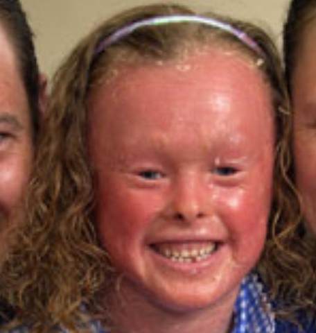
Harlequin Ichthyosis face pictures

Picture 1 – Harlequin Ichthyosis  (face)

Harlequin Ichthyosis Symptoms & Signs

The main symptoms of harlequin ichthyosis are:

Dryness of Skin

The disorder causes dryness of the skin which will lead to scales and dehydration.

Scaly Skin

The disorder causes skin to scale which will lead to having fissure and causes itching and bleeding.

Anatomical Changes

The disorder is known to cause anatomical changes like having poor developed ears and round mouth.

Harlequin Ichthyosis in babies

Infants are the ones who are often triggered and reported of having this very rare skin disorder.

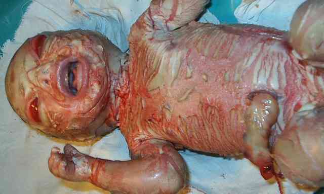
  1. The babies of harlequin ichthyosis are reportedly having a thick, hard skin covering in most part of their body.
  2. The skin has a diamond shaped, medically termed as harlequin, plates which are separated by deep fissures or cracks.
  3. It will greatly affect the shape of the ears, nose, eyelids, and it limits the movement of both legs and arms.
  4. It often pulls the baby’s mouth and makes it widely open.
  5. Aside from that, babies born with this skin disorder have microcephaly or small head and a deformed face.
  6. There will be restriction in the movement of the chest which will lead to difficulties in breathing and eventually will lead to respiratory failure.

Harlequin Ichthyosis Causes

The harlequin ichthyosis is known to be genetic in nature. The ABC 12 gene, which directs the protein production that is necessary to have a normal skin growth, is being mutated which leads to this severe skin disorder. It is a gene that is responsible for the transportation of lipid. When it mutates, it produces a harden protein called keratin. This is an autosomal recessive gene. Autosomal recessive gene means that the parents are carrier of the gene and that they both have one of the genes that they passed on to their baby. One must take note that this occur rarely. That child who doesn’t have the signs of symptoms of this skin disorder will either be a carrier of the gene itself.

The children who are inherited with this form of skin disorder will usually have at birth, a normal skin. They eventually develop roughness and scaling during the first few years of their life. At times it is intermittent. It appears in the infant years and disappears during the adult years, then returns later. It is usually associated with diseases like thyroid, chronic renal failure or cancer.

Harlequin Ichthyosis Diagnosis

Ultrasound to pregnant mother

In diagnosing this type of genetic skin disorder, the mother can undergo ultrasound. It is often the done to be able to diagnose early the baby’s condition prior to the baby’s birth.

Physical examination

Sometimes the diagnosis is made through physically examining your skin and characterizing the scales that is associated with this skin disorder.

Skin Biopsy

If still in doubt, the physician may perform other relevant test such as skin biopsy. This is necessary for him or her to rule out other etiology of the dry, scaly skin that these persons manifests.

Aside from the test that will be conducted, the physician will thoroughly assess you by asking important questions such as any personal or genetic history of ichthyosis, age when the ichthyosis first begun, and presence of other skin disorders.

Harlequin Ichthyosis Treatment

Airway maintainance

In treating this severe and very rare skin disorder, upon delivery, the baby’s airway and circulation is the number one priority. The healthcare team must ensure that the baby or the patient’s airway, breathing and circulation are in stable conditions. The babies will require intravenous access. It will be through this access where the medications will be given. It is necessary to have cannulation of the baby’s umbilicus. The healthcare team will place the babies or infants in an incubator that is humidified. Once the vital signs have stabilized, the baby is then transferred into a level 3 neonatal nursery station or room.

Protection of conjunctiva

It is necessary that the baby will be applied with ophthalmic lubricants in order to protect their conjunctiva. When bathing the baby having this skin disorder, it is advisable that he or she takes a bath twice a day and will use wet sodium chloride compresses frequently. It is then followed by applying bland lubricants to aid in softening the hard skin.Topical keratolytic medications are not to be prescribed in newborns because it has the potential to be toxic to the system.

Intravenous access for feeding

As mentioned, there is a need for intravenous access for intravenous fluids are often prescribed. Neonates diagnosed with this kind of skin disease, initially have feeding problems. There is a need to calculate daily fluid requirements since the neonates will experience excess cutaneous water losses. In addition to that, there is a need for serum electrolyte level monitoring. There is also a high risk of having hypernatremic dehydration. Lastly, there is a need to always maintain a sterile environment to avoid any infection to occur.

Harlequin Ichthyosis Pictures

Harlequin Ichthyosis Pictures in newborn

Picture 2 – Harlequin Ichthyosis in newborn

Harlequin Ichthyosis in infants

Picture 3 – Harlequin Ichthyosis in infants

Harlequin Ichthyosis

Picture 4 – Harlequin Ichthyosis (hand)

No responses yet

« Prev

© 2017 eHealthWall.com. All Rights Reserved. Privacy Policy
This website is for informational purposes only and Is not a substitute for medical advice, diagnosis or treatment.