Cherry Angioma – Pictures, Removal, Causes, Home Treatment
What is Cherry Angioma?
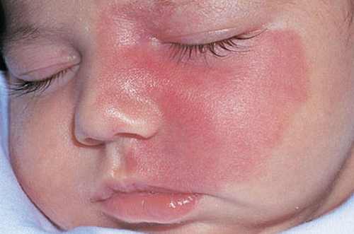
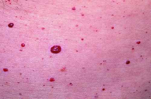
Cherry angiomas are cherry-red eruptions on the skin as a result of proliferation or dilation of blood vessels in the area. Cherry angiomas are the most common kind of angiomas. They are also known as Campbell De Morgan spots owing to the one who discovered it during the nineteenth century, a British surgeon named Campbell De Morgan. These cherry-red papules are also termed as senile angiomas because they occur at old age. They are benign and do not result to malignancies. It may occur on almost all areas of the body, but the most common site are the face and torso.
What causes cherry angioma?
Cherry angiomas usually develop during middle age, but it can occur in younger people. The main cause of the development is rarely understood because people’s interest has not been on it since it does not cause malignancy. A recent study indicates that a defect in the RNA causes the blood vessels and capillaries to proliferate. Specifically, a reduction in microRNA 424 levels is seen in patients with cherry angiomas. This causes increased protein expression and subsequently endothelial proliferation giving rise to dilation of capillaries on the skin. Aside from this, certain risk factors include exposure to chemicals such as butoxyethanol, mustard gas, bromide and cyclosporins. Genetic inheritance also spreads the disease to other people. Stress has also played a role in the formation of cherry angiomas. Emotional, physical and psychological stress leads to faster aging. Development of cherry angiomas increases with age. Cherry angiomas also occur in all races and in both sexes.
What are symptoms of cherry angioma?
Cherry angiomas are made up of groups of dilated capillaries that are evident on the skin. Signs and symptoms include:
- Cherry-red to purple bumps
- Present in the torso, hands, arms, legs, face, scalp and neck
- Young angiomas are the size of a pinhead, but it grows to about .25 inch in diameter
- Spongy, smooth and mushroom shaped
- Painless and harmless
- Bleeding occurs when injured. Cherry angioma on the scalp usually bleeds due to accidental brushing or combing of the hair.
How to Diagnose Cherry Angiom?
Cherry angioma is usually diagnosed with the physical examination of the bump and studying the characteristics. Further tests are not required, but a biopsy may be done to confirm the diagnosis and also helps in the removal of the angioma.
Cherry angioma treatment
Cherry angiomas do not require removal as these are not malignant and they are harmless. Treatment is only considered when they cause frequent hemorrhage or irritation. However, people with cherry angiomas may prefer removal for aesthetic purposes. Minor surgeries are done and medical treatment is not indicated because they do not cause a certain disease and are benign. Patients need to ask their doctors several questions before undergoing treatment to explore the benefits and risks as well as alternatives. Patients may ask:
- What are the methods of removal available?
- Is scar formation possible after treatment?
- What are the risks of undergoing angioma removal?
- Are there any alternatives for surgery?
- Is cherry angioma preventable?
- Does exposure to ultraviolet rays affect the angioma?
- Will angiomas lead to carcinomas or cancer?
- How do we prevent occurrence of cherry angiomas?
Adequate knowledge regarding the angioma may help in decision making and planning for interventions. The following therapies are done for cherry angioma removal:
Electrosurgery or Cautery
Electrosurgery is done by dissecting the angioma using cautery, a special electrical instrument likened to a needle. Scarring is minimal when expert operators are the ones who perform the procedure. Bleeding is also prevented because of rapid closing of bleeders. Local anesthetic is applied prior to the cautery.
Laser Removal
Pulsed dye Laser – Cherry angioma laser removal involves the use of an organic dye mixed with a solvent as the lasing medium. Absorption of the laser energy is possible in the hemoglobin, but this does not cause significant effects.
Intense Pulsed Light (IPL) – IPL is a more advanced method of laser surgery involving more concentrated beam light.
Shave Excision
This involves the delicate removal or slicing of the angioma using a blade in a horizontal angle. Specimen is subjected to biopsy to confirm the diagnosis. Bleeding is prevented through cautery of blood vessels.
Cryotherapy
Cryotherapy or cryosurgery involves the removal of the cherry angioma through freezing by the use of extremely cold substances such as liquid nitrogen (nitrous oxide), carbon dioxide, argon, and dimethyl ether. It also involves destruction, irritation and coagulation. It is a minimally invasive surgery and may result to mild pain that can be reduced by the use of analgesics.
Curettage
Performing curettage is not commonly used like the more conservative therapies discussed earlier. Curettage is the scraping of the angioma using a curette (an instrument used for scraping). Never remove angiomas on your own to prevent profuse bleeding, scarring and infection. Doctors are the ones trained to remove a cherry angioma to arrive with best results.
Natural Remedies for Cherry Angioma
Natural remedies for cherry angiomas are more favored because they are less invasive and less expensive. However, effects may not be seen immediately. Natural remedies are more suited as prevention for cherry angioma formation. The following treatments are the suggested home remedies for cherry angiomas:
Diet modification
Include more fruits and vegetables because these help in increasing the texture and elasticity of the skin. Better skin outcomes mean lesser development of cherry angiomas. Avoid foods that are processed such as canned and junk foods. Include Vitamin A and Vitamin D supplements to promote young and healthy skin.
Topical herbs
Apply sandalwood and basil leaves cream on the cherry angioma to reduce the appearance. These can be pounded and the juice applied on the skin. Witch hazel also reduces bacterial growth, preventing angioma formation
Apple cider vinegar
Traditional remedy includes applying apple cider vinegar to the angioma to dry up the bump. Some individuals who have tries said that the angioma fell off after two weeks of application.
Drink plenty of fluids
Increase water intake to 1.5 to two liters per day. Consume more fluids in the form of juices, jellies and shakes. Good skin hydration prevents angioma and skin breakdown because it enhances the moisture and elasticity.
Avoid stress
Stress is a major risk factor for cherry angioma formation. Learn to do stress reduction activities such as focused breathing, yoga, guided imagery and listening to music. Prevention of stress means prevention of cherry angioma.
Cherry Angioma Prognosis
These cherry-red bumps are most often benign and harmless. It increases in size and number with age, but a sudden development of several angiomas may signal a developing internal malignancy.
Cherry Angioma Complications
Possible complications of angioma include:
- Bleeding when injured, but will easily stop upon pressure application and does not lead to profuse bleeding
- Psychological distress as a result of body image disturbance. These people usually opt to removal of the angioma for aesthetic purposes.
Cherry Angioma Pictures
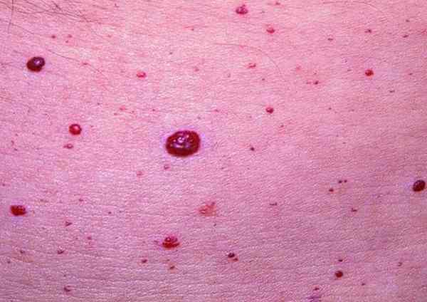
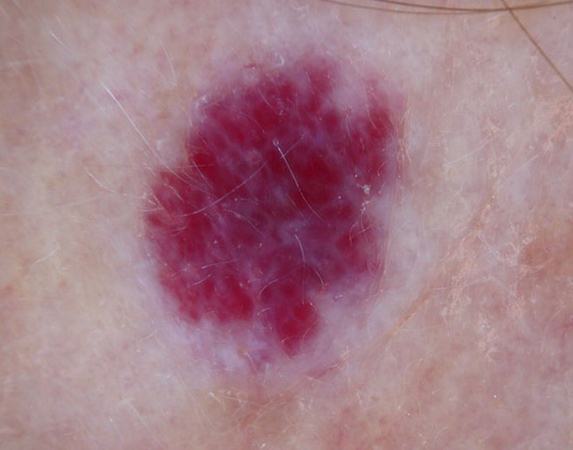
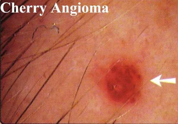
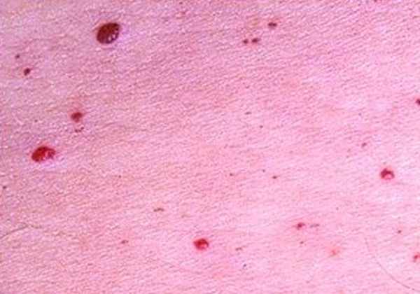
Cherry Angioma on Face
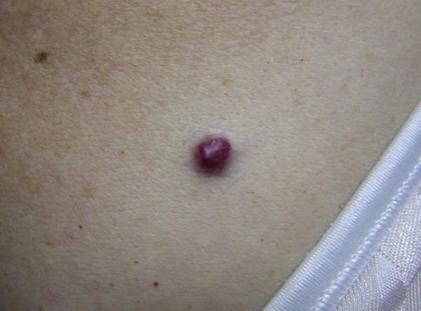
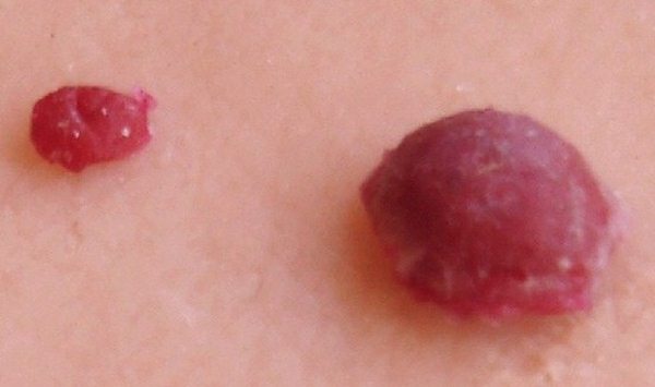
The most common site of blood vessel proliferation is in the face. The vascular lesions are characterized as cherry red to purple in color and flat topped. They usually grow as the individual increase in age and may increase in about few centimeters in size. Some angiomas are close to each other and form a polypoid angioma or a multiple adjoining angioma. These may seem larger and more apparent. Cherry angiomas on the face are sensitive and may cause bleeding when the area is injured. Cherry angiomas on the face also lead to body image disturbance because of the red eruptions on the skin. Although the vascular lesions are harmless, occurrence on the face may result in loss of self-esteem. Removal of the angioma in these people may be considered.
