Spigelian Hernia – Symptoms, Pictures, Location, Anatomy, Causes, Repair
What is Spigelian Hernia?
Spigelian Hernia or Lateral Ventral Hernia is a rare type of hernia that develops through the spigelian fascia. Spigelian fascia is a layer between the rectus abdominis muscle and the semi-lunar line laterally. It is also defined as a protrusion of peritoneal fat or sac of an organ which has a congenital defect or a weakness in the spigelian fascia. It is mostly seen at or below linea arcuata probably because of lack of posterior rectus sheath. It represents approximately 1-2% of all hernia and women are mostly affected and more likely to occur after the age of 50. Oblique hernia symptoms does not show any outward signs like swelling because it penetrates between the muscles of the abdominal wall. This type of hernia is usually small so it’s very difficult to diagnose. Compare to other hernias, because of its size there is a possible risk of strangulation. Predisposing factors that can cause spigelian hernia are increased intra-abdominal pressure, obesity, and trauma.

Abdominal Hernia
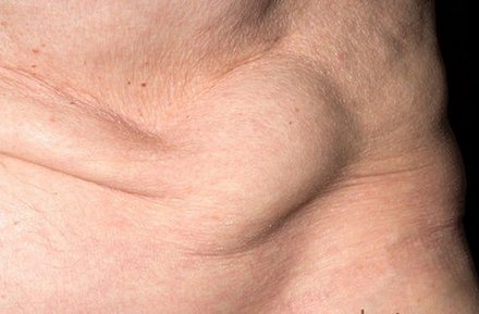
Source – sciencephoto.com
Location and Anatomy of Spigelian Hernia

Picture 2 – Different types of hernia
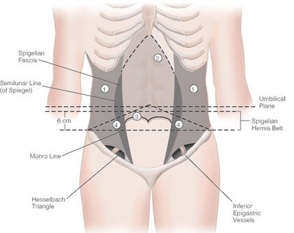
The location of Spigelian hernia is in the lateral edge of rectus muscle and semi-lunar line or below the level of the umbilicus.
Spigelian hernia is a hernia through spigelian fascia, which is the aponeurotic layer that is a layers of flat broad tendons that joins the muscles and it is located between the rectus abdominis muscle or also known as “six pack” medially and the semi-lunar line, it is a curved line placed on either side of rectus abdominis, laterally.
Diagram of Spigelian Hernia
This picture shows the common location of spigelian hernia inside the body.

Spigelian Hernia anatomy and location
Spigelian Hernia Symptoms and Signs
- Localized pain that comes and goes on a repetitive basis.
- Bowel obstruction it’s an obstruction in the intestines, preventing normal transit of products of digestion.
- Nausea and vomiting if there is a presence of bowel obstruction.
These are the only general signs of spigelian hernia. This rare type of hernia is usually small and doesn’t show any outward signs, so, it’s very difficult to determine its presence. The size of this hernia increase the risk for strangulation therefore it’s very necessary to be treated early once it’s detected and confirmed.
Spigelian Hernia Causes
Spigelian hernias usually occur in patients with:
(1) Increased intra-abdominal pressure in situations like having a
- Heavy labor in women can cause pressure in the abdominal area that might result in having spigelian hernia.
- Person who developed urinary retention. Urinary retention is characterized by having a poor, intermittent flow or a sense of incomplete voiding.
- Severe coughing or severe vomiting
- COPD or Chronic Obstructive Pulmonary Disease. COPD is a disease in the lungs in which the airways became narrowed normally due to tobacco smoking which they triggers the inflammatory response in the lung.
(2) Multiparous women or women who gave birth with 2 or more offspring and patients with recent significant weight loss. Clinical findings include focal tenderness or a mass along the linea semilunaris and because of this bowel incarceration and strangulation are common effects that result to spigelian hernia.
(3) Obesity is a medical condition where there is an excess body fat in the body. People that is obese carries an extra weight their muscle are often unable to handle this extra weight which increases the person to have a hernia. A patient that is obese will have a difficulty in determining the location of hernias as well as the recovery of the patient after surgery will become a problem.
Spigelian Hernia Diagnosis
- CT scan or Computed Tomography is considered as a most reliable technique to make sure of the diagnosis. A CT scan uses special x-ray equipment that produces multiple images with different dimension. It helps to identify abnormalities such as tumor and hernia inside the body.

- Ultrasound is recommended as a first line in detecting any presence of mass in the abdomen. Ultrasound is a procedure that uses high frequency sound waves to visualize internal organs and produces images of the body.
- MRI or Magnetic Resonance Imaging is a medical imaging technique that visualize internal organs. MRI can produce a good contrast between the different soft tissue of the body that is really helpful in determining problems in the body.
- Ultrasonography it is used for visualizing subcutaneous body structures such as tendons, muscles, joints, and internal organs.
*These diagnostic exam can help the doctors to detect any unrecognized cases of spigelian hernia. They can show any defect or abnormalities along the spigelian line in the lower abdomen, it can locate the presence of hernia sac, it can show how small the obstruction is and if strangulation is present.
Spigelian Hernia Treatment
There is no specific medical treatment for spigelian hernia because of the risk of strangulation most of the doctors recommend surgery. A surgical procedure in which hernia is repaired or removed and which the abdominal wall is strengthen with surgical mesh. Surgical Mesh is a woven fabric that helps to strengthen tissues, to treat surgical wounds and provide support to organs. Surgical mesh also prevents the hernia to go back.