Archive for September, 2011

Low Grade Squamous Intraepithelial Lesion – Symptoms, Causes, Treatment

Sep 22 2011 Published by under Diseases & Conditions

What is Low Grade Squamous Intraepithelial Lesion (LSIL)?

One cell constitutes a single building block of the body. Everyone already knows that, even little children who have a basic grasp of human anatomy, but due to the current scourge of diseases like cancer, some of these cells that are supposed to build and protect the body have been transformed into ruthless attackers.

So now we have numerous conditions where a specific group of cells are trying to overtake the whole biological system, like acute lymphocytic leukemia for example. There is also a similar disease that is comparable to the former, though Low Grade Squamous Intraepithelial Lesion originates and mainly affects a different part of the body. So now, let us look at the importance of low grade squamous intraepithelial lesion and how it impacts the body of a human being, particularly in the case of women.

Let us start first with the “basics” before getting in to more detail. A squamous cell basically looks like a flat and plate like cell. Usually, this type of cell is more abundant at the edges of the epithelium. The epithelium is a part of the body which covers both the internal and external organs of the body separately.

More often, it also envelops the various parts of the body like the linings of the vessels, body cavities, glands, and organs of different systems and functions. The epithelium also consists of cells which are bound together by some connective material and varies in the number of layers and the kinds of cells.

There are also different types of squamous epithelial cells. There is the simple squamous epithelium, which only has one layer. Examples of this include mesothelium, endothelium, and pulmonary alveoli. The other, which is the stratified squamous epithelium, consists of a basal layer of cuboidal cells and other overlying layers of squamous cells. The epidermis that is a part of our skin, for example, comprises five different layers alone.

Having a low grade squamous intraepithelial lesion means that these otherwise normal cells have some abnormalities in them. The most commonly affected organ, at least for women, is the cervix. The verdict is still out with regards to what causes a low grade squamous intraepithelial lesion. The “culprit” is typically a minor injury that has healed in the cervix. The healing “process” has then caused these squamous cells to exhibit a rapid overgrowth that can only seem as unnatural for the affected patient.

Low Grade Squamous Intraepithelial Lesion Symptoms & Signs

Typically, a low grade squamous intraepithelial lesion will not manifest itself in its early incarnation. However, as the lesion spreads, the different signs and symptoms may appear.

Pruritus

Long-standing pruritus or itching and soreness are the most common initial manifestations of a low grade squamous intraepithelial lesion. Itching occurs in half of all the patients with these lesions.

Bleeding

Bleeding, foul-smelling discharge, and pain may also be present and are usually giveaway signs that the disease has indeed progressed.  There may be some instances when the lesions may be visible and accessible and grow relatively slowly.

Early lesions may appear as chronic dermatitis or skin inflammation. Later on, some of these patients may also notice that a “lump” will then continue to grow and will eventually become a hard, ulcerated, and cauliflower-like in appearance.

Low Grade Squamous Intraepithelial Lesion Causes

As we have mentioned above, the exact cause of a low grade squamous intraepithelial lesion is still unknown. The only thing that most researchers can agree on is the possible risk factors of this condition.

Though a low grade squamous intraepithelial lesion in the cervix does not indicate that the patient is now experiencing cancer, it should serve as a “warning” sign that an occurrence of cancer may happen in the near future.  Low grade squamous intraepithelial lesions represent 4% of all potential gynaecologic malignancies and are seen mostly in postmenopausal women. However, the incidence for younger women is also increasing. The median age for this condition is fifty years of age.

Other possible risk factors that women should be aware of with regards to a low grade squamous intraepithelial lesion are

  1. smoking,
  2. human papillomavirus (HPV) infection,
  3. human immunodeficiency virus (HIV) infection, and
  4. immunosuppression

The less common risk factors are

  1. bartholin’s gland cancer
  2. vulvar sarcoma and
  3. malignant melanoma.

LSIL Diagnosis & Treatment

As much as possible, gynaecologists everywhere are not recommending any kind of treatment to be done with a low grade squamous intraepithelial lesion. The reason is that, unless it has progressed to something far more severe, it should not be attempted to excise or remove the lesion in any way. The best thing that a patient can do is to just “wait”, however impatient patients may be for some drastic procedure to be done.

Gynaecologists or any health care professional is in an ideal position to encourage the patient to perform a vulvar self-examination test regularly using the aid of a mirror. With the mirror, the patient can see what constitutes a normal female anatomy and learn about any changes that should be reported, like the increase in size of the lesion, ulcers, masses, or even a persistent itching. The doctor should urge the patient to seek proper health care if anything abnormal is noticed to be happening inside. A regular pap test every four to six months should also be observed by the patient in question, as well.

However, there may be some cases when a gynaecologist may deem that the lesions may be progressing more rapidly than they had initially expected. In cases like these, some rather aggressive and diagnostic procedures can be employed by the attending physician. One of these is a colposcopy, an examination of the vagina and the cervix with an optical magnifying instrument, which is called a colposcope. A colposcopy is also commonly performed after a pap smear test in the treatment of cervical dysplasia, which is an abnormal tissue development of the uterine cervix. The most dangerous thing about having a cervical dysplasia is that, in about one out of ten cases which have been monitored over recent years, the atypical epithelium which the gynaecologist has observed earlier can progress to a full-blown carcinoma.

A biopsy may also be performed in lieu of a colposcopy. In this procedure, a small piece of living tissue from an organ or other part of the body is removed for microscopic examination. 

The results from this examination can then confirm or establish a diagnosis. It can also estimate the prognosis of the client, or even follow the course of the disease. This kind of procedure is commonly used to determine whether the cancer is present or has spread inside the body.
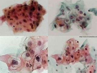
Biopsy results of LSIL under electron microscope.

Picture 1 – Biopsy results of LSIL under electron microscope.

Prevention of LSIL

However, prevention may be as simple as decreasing the incidence of the risk factors associated with low grade squamous intraepithelial lesion. Some simple things, like abstaining from promiscuous sexual activity or having multiple sex partners, can be helpful in reducing the chances of this disease being aggravated.

Read & compare – High Grade Squamous Intraepithelial Lesion (HGSIL) – Symptoms, Causes, Treatment

No responses yet

Side Effects of Aleve

Sep 20 2011 Published by under Drugs & Supplements

Introduction about Aleve

To be fair, Aleve is a really useful drug. One of the many trade names of the naproxen drug, it as a nonsteroidal anti-inflammatory drug (NSAID) which has been used all over the world. Aleve is in the same class of medication as other brand names like Advil or even aspirin.

Aleve is prescribed & used for

  1. relief of fever
  2. headache
  3. migraine
  4. inflammatory symptoms of rheumatoid arthritis (RA) and
  5. osteoarthritis (OA)
  6. relief of mild to moderate pain
  7. treatment of primary dysmenorrhea
  8. ankylosing spondylitis
  9. tendinitis
  10. bursitis and
  11. acute gout

aleve

With so many kinds of disorders and diseases taking, you would think that there could be possibly no wrong with this kind of medication. Well, what most people seem to forget is that Aleve is, first and foremost, functions as a drug. Just like any drug, it can cause some unwanted side effects, and may carry some lifelong complication that may be possibly hazardous to the health of an individual who has taken the drug in question.

Side effects of Aleve

It was really surprising that the severity of the side effects of Aleve is just coming into much scrutiny. For one, taking a regular and prescribed dosage of Aleve may lead to that individual increasing his or her danger for his or her body to develop a heart attack or stroke. Not only that, but there are several well-documented cases of some individuals experiencing nausea and heartburn, something which all  nonsteroidal anti-inflammatory drugs (NSAIDs) also display when the blood levels of a person has reached its toxic “peak”. But the most important thing of all is that clients who are taking Aleve are also putting themselves at risk of haemorrhaging or severe bleeding, since this medication can decrease the body’s natural clotting ability, and thus, can prove to be dangerous if ever these clients have sustained an open injury in any way possible.

Aleve can really be used for just about any health condition out there. Therefore, it may seem to be surprising to some when they have suddenly developed an adverse effect that is related to the medication.

Some of these aleve side effects commonly include a sense of a

  1. vague stomach pain
  2. nausea
  3. vomiting
  4. minor to severe cases of headache
  5. bowel problems like diarrhea and/or constipation and
  6. other sensory problems like drowsiness and dizziness.

There may be times when one of these symptoms can occur “at a flash”, but should one of these continue to endure, then all health care practitioners advise that client to immediately contact the nearest health care official in their town or locality.

There are also other “undocumented” cases with regards to the side effects of Aleve. While some may possibly be minor, other can be more serious in scope and damage. There are also cases of some clients developing major allergic reactions to the drug, while there are those who have developed a severe toxicity to the drug the “side effects” that they are experiencing have proven to be long term in nature. Whatever those may be, all doctors agree that you should have to immediately stop the treatment if you suspect that you have developed one of these side effects while under the therapy of this medication.

No responses yet

Sprained Ankle

Sep 14 2011 Published by under Diseases & Conditions

What is Sprained Ankle?

Sprained ankle is a medical condition that occurs when tough bands of tissues or ligaments tear or stretch resulting to pain and injury. Ligaments are tissues around joints that attach bones together to support and stabilize for movement.

Ankle sprain or sprained ankle can occur when the ligaments are used beyond its capacity causing a momentary stretch usually that happen during physical activity or sudden movement. It is the most common form of orthopedic and soft tissue injury caused by even the simple daily activities such as walking and engaging in competitive, contact sports.

People might interchange the terms ankle sprain and ankle strain, but these terms are entirely different. Ligaments are affected in ankle sprain while muscles are inflicted in ankle strain.

Types

Types of ankle sprain should be known in order to apply the necessary treatment for each, though it is possible that a person canhave a combination of one or more sprains in multiple locations of the ligaments.

Inversion

Inversion also known as lateral sprain and this is the most common type and approximately 90% of the cases have this type. This occurs when the foot is inverted too much causing too much stretch on the lateral ligaments affecting the outside of the foot. This is without joint instability. The majority of the swelling will be mainly around outside the ankles and the location and amount of bruising can be varied.

Eversion

Eversion also known as medial sprain and occurs when there is too much stretch on the medial ligament of the foot. This is without joint instability. This injury is common among high activity and contact sports like hockey, football and basketball. The amount of swelling are not marked though in this case, it can be located inside or near the back of the ankle and causing moderate pain.

High ankle sprain

High ankle sprain medically referred s syndesmotic or syndemosis ligament sprain which occurs when the ligaments that attach tibia and fibula (the two long bones of the lower leg) are injured due to forceful outward twisting of the foot. This type of sprain will took a long time to heal, because of the severity and complexity of the injury. Surgery may be required to help restore and repair the affected ligaments before a thorough rehab program is done.

Classification

There are three classifications of ankle sprain, namely Grade 1, 2 and 3. This is depending on the severity of the damage that the ligaments sustained and the force that is applied to the ankle causing the injury.

Grade 1 – Grade 1 occurs as a mild sprain causing little damage and minor stretching on the ligaments.

Grade 2 – In this grade 2 there is minor tearing of the ligament and the ankle joint is moved awkwardly.

Grade 3 – In Grade 3 there is major tearing of ligament present, presence of instability is felt.

Risk Factors

  1. Participating in sports can cause ankle sprain as much as the body can be vulnerable to intense force and stretching of ligaments especially those are overweight. Unhealthy playing conditions such as uneven surfaces can also increase the risk of sustaining ankle sprain.
  2. Past ankle injuries can also pose a factor increasing the vulnerability of the person to sustain another ankle injury.

Causes

Ligaments can be injured when too much force is used to them and when it is moved beyond its normal range or motion. These are the usual causes of ankle sprain:

  1. Poor ankle flexibility
  2. Weak tendons that cross along the ankle joint
  3. Lax ligaments; can be hereditary or due to overstretching ligaments because of repetitive ankle sprains
  4. Didn’t perform a warm up before physical activities
  5. Failed proprioception on the joints
  6. Slow neuron response to an off-balance postion
  7. Planting the foot on an uneven surface causing the ankle to twist
  8. Losing balance while wearing high heels
  9. Inadequate heel support
  10. When playing contact sports, in the event that one player steps on another causing ankle sprain

Symptoms & Signs

The signs and symptoms of inflammation that will occur on ankle sprain are:

  1. Swelling of the ankles brought about shifting of fluids from inside the tissues.
  2. Pain around the injured area that can be triggered when the affected foot is moved and pressure is exerted on it due to sensitivity of the nerves around the affected area.
  3. The area becomes inflamed and warm due to blood pooling on the affected area.
  4. Decreased or loss of motion

Diagnosis

Diagnosis can be made through complete medical history including the onset of the symptoms. A physical exam will be performed to confirm signs and symptoms. There will be checking for tenderness and the affected area will be tested for different ranges of motion.

When injuries are severe, the physician may request for some diagnostic tests to evaluate more the injury. These diagnostic tests are:

  1. X-ray –  X-ray is the use of small amount of radiation that will pass through your body in order to view the internal structures. This test is good for evaluating presence of fractures or cracks, but less effective in visualizing soft tissue injuries.
  2. Bone scan – Bone scan involves injecting a radioactive material intravenously that will enhance viewing and detecting stress fractures.
  3. Computerized Tomography –  Computerized Tomography scans are effective since it can reveal more detail on injuries because viewing can be made at different angles and can make cross-sectional images of the body’s internal structures.
  4. Magnetic Resonance Imaging – Magnetic Resonance Imaginguses waves and strong magnetic field and is used to visualize soft tissue injuries.

Treatment

Aim of Treatment: To reduce further injury to the ligaments by reducing and keeping swelling, pain and inflammation into a minimum. Sprained ankle management is based on the severity of the injury.

First aid management

For first aid management, it is best to keep the affected area immobilized to prevent further harm to the injured ligaments. A bandage or ankle support may be used to help in immobilization. After immobilization, rest the affected area for 48-72 hours following injury.

Ice applications

Ice applications are also advised 10-30 minutes after injury. Application of less than 10 minutes will have no effect and exceeding 30 minute ice application can damage the skin. It is important to wrap the ice pack in a plastic bag or towel to prevent ice burn. Gentle pressing to the affected part will help reduce blood flow thus limiting pain and inflammation to the site. After the first application, physicians recommend re-applying of cold packs for 15 minutes every two hours for the first 48-72 hours of injury to achieve the maximum desired effect.

Compression with the use of a bandage

Compression with the use of a bandage will help limit swelling. It is important that mild pressure is applied that is to not to impede the blood flow.  Before going to sleep, it is important to remove the bandage. After 48 hours following injury, it is indicated to remove the bandage because it may limit the movement of which the ankle should be moving freely and free from pain. However, there are people who keep their bandages longer to keep their joint more comfortable and decrease further swelling.

Elevation is done to limit swelling. It is advised to keep foot up on a chair at hip level when sitting and when lying on bed, a pillow can be placed under the affected area.

Contraindications

Application of heat within 72 hours after injury would increase the blood flow to the affected area when presently inflammation is developing, so it must be avoided. However, application of heat after 72 hours following injury, heat can be soothing to the affected area.

Ingestion of alcoholic drinks are also must be avoided because this will increase bleeding and swelling thus decreases fast healing.

Further movement may also aggravate the condition and massage within the 72 hours of injury can increase swelling. However, as with heat, massages can be soothing to the area affected.

Other Treatments

Wearing of ankle support while doing daily activities until symptoms are gone can be used to support the healing ligaments. Physicians also suggest that to keep all of the weight on your ankle, use of crutches and walkers can be considered. It also advised to refrain from sports 3-4 weeks after the injury to promote further healing of the torn ligaments.

Uses of medications like over the counter pain relievers such NSAIDs or nonsteroidal anti-inflammatory drugs can relieve pain and limit swelling and inflammation. It is also important to watch out for side-effects of these drugs such as stomach pain and bleeding pain. That is why these medications should be taken after meals to avoid gastrointestinal irritation. Some doctors say that they don’t recommend taking anti-inflammatory painkillers for the first 48 hours because this may delay healing. They implicate that inflammation is a part of the healing process and taking in these painkillers might disrupt it. Use of topical anti-inflammatory painkillers can also be used in treating sprains and there is less risk for side-effects.

Rehabilitation

Rehabilitation exercises for ankle sprains can be done and performed at home to prevent chronic pain and promote healing. An individual can perform stretching exercises before and after physical and vigorous exercises to prevent re-injury.

Ranges of motion exercises are also important for rehabilitation. This involves performing a set of exercises with repetition of 10-30 times and this set is done 3 to 5 times a day. An example of which, is tracing the alphabet with your toe which encourages all directions of ankle movement. Another is sitting on a chair with foot flat on the floor and moving the knee side to side slowly while maintaining the foot flat.

Towel curls can also be performed. This is done by placing a hand towel on a smooth floor and curling the toes around the towel to grab it and continuing to scrunch the entire length of the towel.

Strengthening exercises can be performed when an individual is able to stand without feeling of increased pain. These exercises should be held for at least 3 to 5 seconds with 15-20 repetitions once or twice a day for 2 to 4 weeks. It is done by starting sitting on the floor with fool flat on it and pushing it outward against an immovable object such as a wall.

Complications

If sprained ankle is left untreated or if an individual engaged in an activity, one may experience the following complications:

  1. Chronic pain
  2. Early onset arthritis
  3. Chronic ankle joint instability

Prevention

In order to prevent occurrence or sprained ankle, these are the following steps:

  1. Warm up before engaging in exercise and playing sports
  2. Be cautious in walking, running on an uneven surfaces
  3. Use footwear that are comfortable and made for your activity
  4. Be careful when wearing high heels
  5. If you are not fit or conditioned for a certain type of sport, don’t engage in it.
  6. Practice balancing exercises – Perform exercises that promote muscle strength and flexibility.

Pictures

Sprained Ankle Pictures

Pictures of Sprained Ankle

Sprained Ankle Pictures

Image of Clinical presentation of Sprained Ankle

Sprained Ankle Pictures

Photo of Swelling and redness in Sprained Ankle

Sprained Ankle Pictures

Support band pic in Sprained Ankle

No responses yet

Churg-Strauss Syndrome

Sep 12 2011 Published by under Syndromes

What is Churg-Strauss Syndrome?

Churg-Strauss syndrome is a health problem characterized by swelling of the blood vessels. This swelling can impede blood circulation to essential body organs and at times may lead to irreversible damage. The most usual manifestation of Churg-Strauss syndrome is asthma. However, it can also result to different problems scaling from allergic rhinitis, skin rash and gastrointestinal bleeding, to terrible pain and lack of sensation in your hands and feet. The broad variety of symptoms and their likeness to manifestations of other diseases make it more difficult for doctors to diagnose Churg-Strauss syndrome. Churg-Strauss syndrome is uncommon and has unknown cure at present. However, your physician can typically assist you in managing symptoms by means of steroids and other immunosuppressant medications.

Symptoms & Signs

Churg-Strauss is an extremely unpredictable disorder. Several patients manifest merely minor symptoms, while some suffer major or critical problems. Presently, there are three stages of Churg-Strauss syndrome, everyone having its individual manifestations, although not everybody will undergo the entire three stages or in the similar arrangement. This is particularly correct as soon as the illness is diagnoses and managed earlier than the most severe injury happens.

churg strauss syndrome pictures

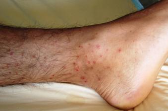
Pictures of symptoms of churg strauss syndrome (allergic, vasculitic stage)

Churg-Strauss stages comprises of:

Allergic stage

This is more often than not the initial stage of Churg-Strauss syndrome. It’s characterized by some allergic reactions, involving:

Asthma – The most usual manifestation of Churg-Strauss syndrome, asthma forms on standard three to nine years prior to the appearance of other signs and symptoms. In patients who have Churg-Strauss syndrome who previously suffer from asthma, symptoms typically grow to be bad and might necessitate steroids for regulation. Some people have what is called as late-onset asthma. Having asthma, even later on in life, doesn’t essentially signify that you obtain Churg-Strauss syndrome, nevertheless, Churg-Strauss happens hardly ever as a result of asthma.

Allergic rhinitis – This involves your nose’ mucous membranes, resulting to runny nose, sneezing and irritation.

Sinusitis – You may suffer from pain in the facial area and have nasal polyps, which are soft, non-cancerous lumps that brought about by inflammation for a long period of time.

Eosinophilic stage

An eosinophil is a kind of white blood cell that facilitates protection from particular infections. Usually, eosinophils comprises only an insignificant percentage of white blood cells, although in Churg-Strauss syndrome, unusually these cells are elevated medically termed as hypereosinophilia. Hypereosinophilia can lead to severe damage. Signs and symptoms of hypereosinophilia will be based on which area of your body is involved. The most targeted areas are your lungs and digestive tract. Hypereosinophilia can signify a quantity of allergic reactions and doesn’t essentially denote that you will have Churg-Strauss syndrome. Generally speaking, signs and symptoms of the hypereosinophilia stage may involve:

  1. Fever
  2. Loss of weight
  3. Asthma
  4. Tiredness
  5. Sweating at night
  6. Cough
  7. Pain in the abdomen
  8. Abdominal bleeding

This stage can continue in months or years, and your manifestations may vanish sometimes, simply to come back afterward. You might also suffer symptoms of hypereosinophilia and systemic vasculitis, the third stage of this disorder, all at once.

Vasculitic stage

The characteristic sign of this stage is massive blood vessel swelling medically termed as vasculitis. Through thinning blood vessels, swelling lessens blood circulation to essential organs all over the body, together with your skin, heart, peripheral nervous system, muscles, bones and digestive tract. Seldom, your kidneys might also be involved. Throughout this stage, you may experience being sick and have unintentional weight loss, inflamed lymph nodes, feebleness and exhaustion. Based on which areas are involved, you may also suffer from:

  1. Rash or sores in the skin
  2. Joint pains and inflammation
  3. Intense pain, loss of sensation and tingling in your hands and feet medically known as peripheral neuropathy
  4. Intense abdominal pain
  5. Diarrhea, nausea and vomiting
  6. Difficulty of breathing from asthma or congestive heart failure
  7. Coughing up of blood
  8. Chest pain
  9. Irregularities in your heart rhythm
  10. Blood in your urine

It is recommended to consult your doctor any time you manifest signs and symptoms familiar to Churg-Strauss syndrome. Churg-Strauss syndrome is uncommon, and it’s more possible that these manifestations have some other origin. However, it’s significant that your physician assess them.

Treatment

At present, the cure for Churg-Strauss syndrome remains unknown. Although specific drugs may aid even patients with grave symptoms attain remission. An excellent result and a decreased chance of complications from both the disorder and its management are greatly possible when Churg-Strauss syndrome is confirmed and managed ahead of time. Drugs indicated to manage Churg-Strauss syndrome involve:

Corticosteroids

Prednisone is the most usually given drug for Churg-Strauss syndrome. Your physician might give a high dose of corticosteroids or an increase in your present dose of corticosteroids to get hold of your symptoms as quickly as possible. However, since high doses of corticosteroids might lead to severe side-effects, such as bone thinning, elevated blood sugar, cataracts and infections, your physician will reduce the dose slowly until you are getting the minimum quantity that will maintain your disorder under management.

Other immunosuppressive drugs

Indicated for people with minor symptoms, a corticosteroid alone may be sufficient. Some people may need one more immunosuppressive drug, such as Cytoxan, Imuran or methotrexate, to decrease the body’s immune response even more. Since these medications damage your body’s capability to battle off infection and might lead to other grave side-effects, your disorder will be personally observed while you’re having them.

Intravenous immunoglobulin or IVIg

The infusion is given per month. IVIg is the most gentle of the medications prescribed for Churg-Strauss syndrome. The most usual side-effects are flu-like manifestations that normally persist just a day or so. IVIg has two most important disadvantages, on the other hand, it’s very costly, and it isn’t generally successful. IVIg isn’t regarded as an initial management for Churg-Strauss syndrome, however, research have revealed that it can be useful for patients who don’t react to other drugs. Even though pharmacologic management can alleviate symptoms of Churg-Strauss syndrome and propel the disorder toward remission, relapses are still frequent.

Prognosis

Churg-Strauss syndrome is a grave disorder that can be life-threatening. Not treated it is tremendously hazardous and jeopardizes the organs that are involved. With forceful management and close observing it might be calmed and remission is likely.

No responses yet

Tumor Lysis Syndrome

Sep 11 2011 Published by under Syndromes

What is Tumor Lysis Syndrome?

Tumor lysis syndrome is a serious metabolic crisis often linked among particular forms of tumors. Dilutions of electrolytes inside the cell, vary from electrolytes outside the cell. In tumor lysis syndrome, tumor cells break apart, liberating their insides into the blood vessel. It will lead to a hazardous modification in the usual equilibrium of fluid and electrolytes—the amounts of potassium, phosphate and uric acid are increased, whereas calcium levels are reduced. The alterations happen in a very rapid manner and may possibly be so fast that instant fatality can occur.

Symptoms & Signs

Initially patients are asymptomatic or they do not manifest any symptoms, however, as the syndrome progresses it will lead into the following symptoms:

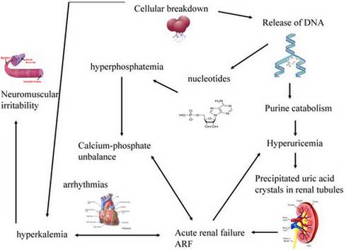
Tumor lysis syndrome pathophysiology, symptoms picture

Picture of Tumor lysis syndrome pathophysiology and symptoms

  1. Heartbeat irregularities
  2. Damage to mental ability
  3. Loss of awareness
  4. Difficulty of breathing
  5. Elevated blood potassium and uric acid levels shown in the laboratory tests
  6. Decreased blood calcium levels also shown in the laboratory tests

Treatment

Treatment is intended at preventive and palliative care, with the primary objectives to avoid kidney impairment and serious electrolyte problems. Patients who are vulnerable obtain treatment on a hospital to permit critical monitoring by hospital staff. The patients must have access to a dependable intravenous line always. Before starting treatment, a patient’s fluid levels and electrolyte status are cautiously calculated and examined. If the results came out and there are irregularities, the doctors may consider holding the treatment for a while, although this is not all the time a choice.

Laboratory examinations are completed regularly to check serum calcium, potassium, phosphate, magnesium and uric acid levels. A normal hospital procedure may need blood be removed for these examinations every two to six hours throughout two to three days. Subsequent are preventions and treatment plans for every major electrolyte problems like hyperuricemia, hyperkalemia, hyperphosphatemia, and hypocalcemia.

Hyperuricemia

Hyperuricemia is defined as an irregular elevation of serum uric acid levels that may possibly result to acute renal failure. There are a number of ways available to avoid renal impairment lie forceful hydration for instance. Also, drugs labeled as diuretics, for example furosemide or acetazolamide, are prescribed to aid increase urine excretion when needed.

Prophylactic allopurinol can also be prescribed to patients vulnerable for tumor lysis syndrome. One dose of 600 mg can be prescribed the day prior to treatment, followed by 300 mg once daily for the rest of treatment. Allopurinol is helpful since it stops the uric acid formation.

Hyperkalemia

Hyperkalemia is defined as an irregular elevation of serum potassium levels that may possibly result to hazardous irregularities in heart beats, heart attack, and muscle weakness. Regular checking with electrocardiography is suggested in patients vulnerable for tumor lysis syndrome in order that changes in the electrical movement of the heart can be detected ahead of time. Foods rich in potassium can also be limited to avoid by now high levels from elevating. Every now and then, drugs like Kayexalate are prescribed to facilitate reduction of serum potassium levels.

Hyperphosphatemia

Hyperphosphatemia is defined as an irregular elevation of serum phosphate levels that may possibly result to neuromuscular irritation and deteriorate kidney function. Cancer cells can have up to four times as much phosphate as non-malignant cells. Patients having acute tumor lysis syndrome can be taught to lessen their food intake of phosphate. Additionally, they can be prescribed drugs that attach to phosphate, thus preventing its intestinal absorption.

Hypocalcemia 

Hypocalcemia is defined as an irregular reduction in serum calcium levels that may possibly result to tetany, muscle cramps, and seizures. A calcium supplement can be prescribed.

Dialysis

Dialysis is a process utilized to control electrolyte problems by means of diffusion and ultrafiltration of fluid. Hemodialysis is a process that eliminates waste products via the human’s blood. Dialysis can otherwise be executed via the peritoneum called peritoneal dialysis. Since peritoneal dialysis does not clean up phosphate and urate as proficiently, and since it is not possible in patients with abdominal growths, hemodialysis is the ideal process. A physician who focuses in nephrology will normally inspect a vulnerable patient prior to cancer management, to get ready for the option of dialysis. In several cases, dialysis begins as a precautionary measure, both prior to or throughout cancer treatment.

No responses yet

Locked-in syndrome

Sep 10 2011 Published by under Syndromes

What is Locked-in Syndrome?

Locked-in syndrome is an uncommon neurological problem described as full paralysis of voluntary muscles in the entire area of the body with the exception of eye movement that the individual can still control. It may possibly the consequence of brain injury, circulatory system diseases, diseases that tear down the myelin sheath covering the nerve cells, or drug overdose. Patients with locked-in syndrome are aware and are still capable of thinking and reasoning, but incapable of speaking or moving. The problem will lead individuals to turn out to be absolutely mute and paralyzed. Communication may be doable with blinking and eye movements.

Having a locked-in syndrome is really difficult for the individual and with his or her family. Especially that the patient will become bed ridden and will be incapable of speaking. Despite the problem the family should just have to take into consideration that the individual is still aware of his or her surrounding and can still think and reason. He or she may communicate using blinking and eye movements. In some cases, numerous therapies shows that function can still be slowly regained.

Signs & Symptoms

Symptoms of locked-in syndrome include the following:

Locked-in syndrome pictures

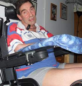
Picture of voluntary muscle dysfunction in Locked-in syndrome

  1. Full incapability for voluntary muscle control except eye movements and blinking
  2. The patient can still think, reason and remain conscious and aware
  3. Usual sleep and wake cycles are preserved
  4. Not capable of speaking
  5. Paralysis of lower extremities, lower cranial nerves and voluntary muscles

Due to the following symptoms of locked-in syndrome the patient required full care in a hospital or in a home setting.

Causes

The common causes of locked-in syndrome are the following:

  1. Traumatic brain injuries caused by stroke, severe head injury or ruptured aneurysm
  2. Diseases that tear down the myelin sheath surrounding the nerves, drug toxicity
  3. Any neurologic problem that can involve the ventral pons in the brain, which is the one responsible for voluntary muscle control

Treatment

As of now there is no known cure or surgery for locked-in syndrome. The management for the problem is only supportive care. They are taken care of critical care specialists, doctors of neurology, and physiatrists. They can also be supported by

  1. physical therapists
  2. occupational therapists
  3. speech and language therapists and
  4. psychotherapists.

Prognosis

The prognosis for locked-in syndrome is poor because there is no known cure and no standard treatment. In some cases therapy can help patients to regain their function. Despite the general poor prognosis there are still cases of locked-in syndrome who have regained impressive improvement of function with the help of new technologies like augmentative communication technology.

Now that there are new hopes for locked-in syndrome, families of the patients should not give up and maybe sometime in the future therapy could really help majority of the patients for their improvement and slowly return to their productive life.

No responses yet

Next »

© 2017 eHealthWall.com. All Rights Reserved. Privacy Policy
This website is for informational purposes only and Is not a substitute for medical advice, diagnosis or treatment.