Archive for September, 2011

Prognosis of Liver Cancer

Sep 10 2011 Published by under Diseases & Conditions

Liver cancer occurs due to metastatic effect of other cancers on liver. Prognosis of liver cancer depends on the stage of cancer. If the cancer is in the early stage, its prognosis is good whilst if is in the advanced stage, the prognosis is poor. The habit of smoking in the patient has also the capability of judging the prognosis level of lung cancer. The prognosis of liver cancer depends on the extent of metastasis, size and number of the tumors, functioning of the liver, levels of alpha-fetoprotein (AFP) and general body health.

Prognosis of secondary (metastatic) liver cancer

Metastatic liver can be diagnosed when patient exhibits tenderness and pain in the liver. Metastatic type of liver cancer is very dreadful and fatal. Prognosis of this is poor. Patients once diagnosed with metastatic liver cancer have a maximum life expectancy of less than a year.

Prognosis of stage 4, 3, 2 of liver cancer

Stage two of liver cancer is determined by small sized nodules or tumors within the liver. Treatment is focused on either killing the cancer cells by treatment or atleast stopping further growth of the tumors. Prognosis of this stage is comparatively good.

Stage three of liver cancer is determined by the spread of the tumor to other specific organs such as lymphatic tissues, unilaterally on both sides of diaphragm. This condition is known to be advanced locally. Prognosis of this stage depends on the extent of local organs involved and the extent of lymphatic tissue damage too. Prognosis is fair if the cancer can be eliminated or atleast prevented from further growth.

Stage four of liver cancer is fatal as it advances to several body organs like lungs, gall bladder, bones and even brain too. Prognosis of this stage is poor. The life expectancy of the patient will also be reduced to not more than one month.

Life expectancy of patients with liver cancer

If liver cancer is detected in early stages like stage 1, treatment options to cure the disease are more. In such cases the life expectancy of the patient will not be affected much. In cases of lung cancer in stage 2 or 3, the disease is advanced locally and hence with proper care or treatment the life expectancy of the patient can be 5 years and if the mile stone is crosses it can extend to 30% more. In cases where there is liver transplantation done, about 75% cases achieve a life expectancy of 5 years. In metastatic/ 4th stage of liver cancer, transplantation of liver has no use and the expectancy rate is just about I year.

Prognosis of liver cancer without treatment

In severe cases, i.e. stage 4 cancer treatment will be of no use. Liver transplantation will have 0% effect on the patient. Prognosis of liver cancer without treatment is generally poor.

On the whole, the prognosis of liver cancer varies from person to person and also depends on the stages of cancer.

No responses yet

Periorbital Cellulitis – Symptoms, Causes, Treatment & Home Remedies

Sep 09 2011 Published by under Diseases & Conditions

What is Periorbital Cellulitis?

Periorbital cellulitis occurs when the eyelids and skin that surround the eyes are infected. This is due to infection made by pathogenic microorganisms that enter in the breaks of skin in the eye. It also doesn’t travel from an infected eye to the other. This medical condition affects children under the age of six. However, the inflammation can spread to the nasal sinuses causing sinusitis. A child’s risk for periorbital cellulitis can be increased when deep tooth and mouth infections are present, there is presence of styes or eyelid bumps and the lacrimal gland (the gland that produces tears) is swelling.

Periorbital cellulitis should not be confused with orbital cellulitis. Orbital cellulitis is a more serious problem and can be a life-threatening condition. The infection on orbital cellulitis is found on the septum of the orbit considering it as an ophthalmic emergency.

There are two different kinds of periorbital cellulitis. It depends on the soft tissue infected. These are:

  1. Preseptal cellulitis – this involves the eyelids and the tissues around the eye, but not affecting the eyeball. This type of infection can spread to different parts of the face but the orbital septum, the membrane that separates the orbit from the facial soft tissues, helps keep the orbit from being affected.
  2. Preseptal periorbital cellulitis – this is less serious since it doesn’t affect the eye and socket but it can spread to the orbit and it is always seen on small children.

Causes

There are different causes of periorbital cellulitis:

  1. Skin trauma or a break or laceration in the skin
  2. Infection brought about a bug or insect bite
  3. Spread of infection from paranasal sinuses or dacryocystitis
  4. Infection from the respiratory tract

Common causative agents in periorbital cellulitis are Staphylococcus epidermidis, Staphylococcus Aureus, Haemophilus Influenzae, Steptococcus and some anaerobic microorganisms.

Symptoms & Signs

Children can’t tell if they are having abnormalities in their body, therefore it is up to the parents to notice deviations from their children. Parents should see and observe marked changes on the face and in the eyes of the child. The symptoms manifested are:

  • Tenderness around the affected area
  • Swelling of eye lids
  • Conjunctivitis or pink eye
  • Low grade fever
  • Only one eye is affected
  • The child had difficulty in opening their affected eye lid
  • There are red streaks on the skin of the eye
  • The child becomes agitated
  • The child has colds
  • In some cases there would be presence of drainage coming from the eye
  • The child’s eye movements and able to see are not affected

Immediate help is needed when:

  • The child reports blurred or double vision
  • The child reports photosensitivity
  • The child experiences high fever and chills
  • The child reports that his or her neck feels stiff and is unable to move it
  • The child experiences seizure

Diagnosis

When the patient is brought to the hospital, an ophthalmologist will determine if the patient was suspected with cellulitis in the eye area. During this consultation, the patient’s medical history will be taken to determine the origin of the infection, recent sinus infection and dental problems. Physical exam will also be done to check deviations and abnormalities in the affected area. The following laboratory and diagnostic tests may also be needed to diagnose periorbital cellulitis

Blood Tests – This is done to determine what type and what kind of bacteria causes the infection.
Drainage culture – Same as with blood tests, this is done to determine the pathogen involved

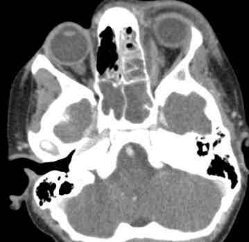
Computed Tomography scan

Computed Tomography scan test will visualize your child’s eye structures such as bones, muscles and blood vessels around the eyes. A dye can be introduced to help in visualization. It is important to check first the child’s history of allergies to seafood and iodine to prevent anaphylactic reactions.

Treatment

Periorbital cellulitis treatment regimen includes:

  • Giving of antibiotics for periorbital cellulitis.

For mild cases, oral antibiotics are prescribed to kill the pathogenic microorganism and prevent complications. Some of the antibiotics prescribed are: penicillin and co-amoxiclav in which it may be required to be taken seven to ten days for adults and for children affected, they may be asked to take it in three divided doses for twenty-four hours.

When cases are severe, the patient will be admitted to the hospital and IV antibiotics will be given two to three days. A drug which usually prescribed is Ceftriaxone 2mg of injection. Once the treatment is finished, the patient’s antibiotics will return into oral form to prevent the disease from recurring.

  • Anti-inflammatory drugs are also given to reduce the child’s pain and inflammation such as ibuprofen.
  • Medications for colds and eye drops or ointments are also prescribed.
  • Tetanus vaccines are also given to prevent tetanus, a life-threatening infection because your child can acquire it from deep cuts and puncture wounds.
  • Warm, moist compresses can also help soothe the affected area.
  • For cases where there is lid abscess or sty, an eye specialist is needed to drain it.

To determine the effect of the treatment, the physician may require a follow-up visit, a week or a two later in which the infection should be long gone. This infection may clear up as little as 48 hours.

It is also important for the child to wear safety equipments to protect his or her face from injury. This is to guard the face from lacerations and trauma. In the event that a wound is present, it is advised to clean the affected area and keep it dry. The use of soap and water would be enough and covering the wounds with dry bandage will complete it. This is to prevent bacteria from harboring opened skin wounds.

Home Remedies

For adults, home remedies are suggested since they can determine which type is actually effective on them.

  • The use of coriander seeds to use it to wash the eyes and face are essential to eye infection relief.
  • Warm compresses of rose oil, lavender and chamomile help soothe the affected area.
  • Oils with antibacterial properties such as tee tree oil can produce a faster recovery from the infection.
  • Soaking of cloth into aloe vera juice will also help relax the infected eye.

Is Periorbital Cellulitis Contagious?

The periorbital cellulitis itself is not contagious, but the bacteria that cause periorbital cellulutis can cause other infection especially if it goes through the bloodstream.

Pictures

Periorbital Cellulitis eye normal eye pictures

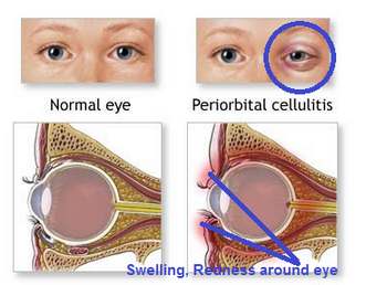
Picture of normal eye and periorbital cellulitis eye comparison

Periorbital Cellulitis Images in Children

Picture of swelling, redness in children of periorbital cellulitis patients

Periorbital Cellulitis Images in women teenagers

Periorbital Cellulitis Image in women teenager (right eye)

Periorbital Cellulitis demonstration

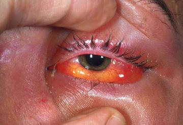
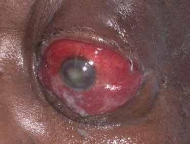
Periorbital cellulitis diagnosis (look at the redness and odema of eye)

purulent discharge from periorbital cellulitis eye picture

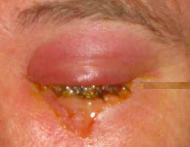
Purulent discharge from periorbital cellulitis eye

severe condition of periorbital cellulitis picture

Severe form of periorbital cellulitis

ct scan showing periorbital cellulitis image

CT scan (brain) showing periorbital cellulitis

No responses yet

Mucositis – Definition, Symptoms, Grading, Pictures and Treatment

Sep 07 2011 Published by under Diseases & Conditions

What is Mucositis?

Clients undergoing chemotherapy for cancer frequently have mucositis (sores in mucous membranes) of the entire GI tract, especially in the mouth (stomatitis). Normally, the mucous membrane of the tract undergoes rapid cell division and replaces dead damaged cells quickly. In chemotherapy, mucous membrane cells are killed more rapidly than they are replaced resulting in sore formation. An individual may have a sensation a bit like getting burn in the throat. Mouth sores are painful and interfere with clients’ desire and ability to eat. The patients most likely may require medication for pain for a period of one week or two until the tissues get better.

Mucositis is the effect of modern cell-killing treatment for cancer patients. It is in addition the chief source of pain for patients who go through transplants for the bone marrow.

Symptoms & Signs

Mucositis because it is an inflammation of the mucous membranes it includes the symptoms of redness, pain and ulceration in the affected area.

Aside from the main symptoms, there are also numerous outcomes that may take place due to the occurrence of mucositis and these include.

  1. Decrease in the volume of blood (Hypovolemia)
  2. Fluid and electrolyte imbalances
  3. Malnutrition (underweight)

If the patient is not taken care of, these complications are possible to be deadly based on the extent of severity.

What makes it more complicated is that chemotherapy usually cause nausea and vomiting and what happens is that the mucous coating the inside of the mouth are predominantly susceptible and at risk to contamination and ulceration. Furthermore, vomiting can cause the mouth’s lining and the gastrointestinal tract to become aggravated. The damaged cells in the mucosal lining will be incapable of healing normally.

An additional effect of this condition is the loss of taste. This adds to the amount of the power and assertiveness of the chemotherapy that the patient is having. Due to the mouth sores, eating will be very difficult and will be even harder if you are unable to taste the food you are eating. If you cannot eat properly you will start to have weight loss and eventually will also lose the integrity of the muscles. They will have deficiency in energy and therefore will have a weakened immunity.

Causes & Risk Factors

1. Chemotherapy

  1. Can get in the way of eating, drinking, and living
  2. Can be a reason for an increased risk of infection, reduction of appetite, and interruption in cancer management
  3. Will make the person with mucositis miserable

Sores can appear five to ten days after chemotherapy starts and their level of severity and occurrence will be based on the particular chemotherapy

Frequently, patients require high doses of chemotherapeutic agents, or combined chemotherapy and radiation for stem cell or bone marrow transplants preparation. Unfortunately, such high doses clean out the bone marrow and this amplify the probability of mouth sores

Chemotrapeutic drugs such as Doxorubicin, or vincristine, 5-FU and methotrexate, or taxoids like docetaxel frequently cause mucositis

2. Radiation

Radiation in the head or neck area can cause mucositis

Both of them show aggression in cells for the period of their separation process. Therefore, cells that separate quickly, like those in the lining of the mouth which separates every three days are frequently involve. This can produce mouth sores since the cells that are assaulted die and do not rejuvenate. When they don’t rejuvenate, the result will be mouth ulcers. Also, a reduced white blood count can make the sores emerge more commonly and also strengthen the risk of infection.

Grading

World Health Organization’s Oral Toxicity Scale

Grade 1 – Pain and redness

Grade 2 – Redness, ulcers, swallowing solid food is possible

Grade 3 – Ulcers with widespread redness; swallowing solid food is not possible

Grade 4 – Mucositis to the degree that receiving nourishment is not possible

Treatment

Infection control

In treating mucositis experts agree that to lessen the infection brought about by mouth sores keeping good mouth hygiene is still the best way.

The cleaning of the mouth is very important. Infections make the lives of patients undergoing chemotherapy and radiation difficult. Given that the mouth in nature shelters microorganisms like bacteria and fungi, it’s vital to maintain the mouth’s hygiene in an attempt to avoid contaminations.

The regularity of treatment is the most principal aspect in decreasing the gravity of disease and in upholding the healing of the ulcers. It is advisable to clean the teeth and mouth every four hours or after each meal and before going to sleep.

It is recommended to brush the teeth softly and to pay careful attention on your gum line. Brush two to three times daily using soft, bristled brush made up of nylon. If they don’t cause distress to the mouth, electric and ultrasonic brushes may be used. Foam brush or piece of gauze is more preferred by some health professionals who say that it’s safer to not use a brush to clean the mouth. The gauze can be washed in water with salt or prescribed mouthwash. In cases wherein there’s a low platelet count (fewer than 20,000) or low absolute neutrophil count (fewer than 500) it is necessary to use either the foam brush or gauze instead.

When a pediatric client used floss it is recommended to use waxed or taped floss to aid in the prevention of further damage to soft tissue, but it is absolutely necessary for the child to not floss if his ANC is 500 or less.

Secondary infection control

To avoid secondary infection the doctor may prescribe antivirals and antibacterials such as Nystatin, Acyclovir, Clotrimazole, Fluconazole, etc.

Mucositis caused by Radiation or chemotherapy

At present there is no recognized way of avoiding mouth sores caused by chemotherapy or radiation.

Pictures
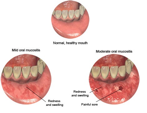
mucositis picture - redness, swelling, sore of mucus memebrane

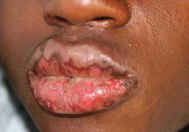
Mucositis picture – redness, swelling, sore of mucus membrane of lips
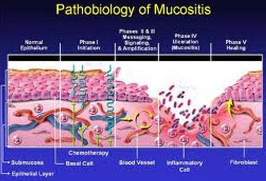
mucosistis pathobiology picture

Image of Pathobiology of mucositis

Image of Mucositis of lips in teenagers
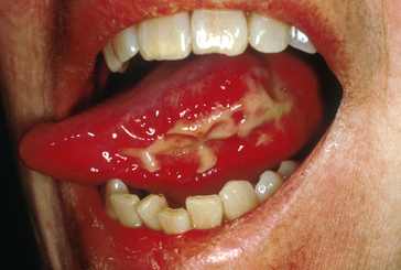
Image of Mucositis of lips in teenagers

mucositis picture showing redness, swelling, ulcer of mucus membrane

Mucositis picture showing redness, swelling, ulcer of mucus membrane

Mucositis Pictures

Picture of grade 2 mucositis

Mucositis Pictures

Picture of Grade 4 mucositis

No responses yet

Fuchs Corneal Dystrophy – Symptoms, Treatment and Surgery

Sep 05 2011 Published by under Diseases & Conditions

What is Fuchs Corneal Dystrophy?

Fuchs Dystrophy is a disease that involves the eye’s cornea. It is a condition that can be hereditary. The cornea is the transparent layer at the front of the eye that helps focus rays of light. It is very common that the disease includes both of the patient’s eyes and a little more frequent in females than males.

Pathophysiology

So now that we know the definition of this disease, we now need to know how this disease happens. The endothelial cells or the cells that compose the posterior layer of the eye’s cornea start to weaken and later on this will result to improper functioning. And then these cells little by little gradually will not be able to compensate. The cornea will now become condensed with additional fluid. In the later stage, the manifestations like for instance poor vision and pain in the eye may develop.

The treatment will vary from patient to patient. Some patient can be treated effectively with the use of prescribed ointments and eye drops. However, some patient may need a surgical transplant of the cornea.

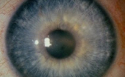
Fuchs corneal dystrophy

Symptoms & Signs

Some physicians may already perceive early signs of Fuchs’ Endothelial Dystrophy in 30 years old and 40 years old patients. Nevertheless most patients are asymptomatic until they reach the age of 50 and 60, in which they start to manifest the symptoms.

Signs and symptoms of Fuchs’ Dystrophy include the following:

  1. It commonly involves both eyes   
  2. Poor/Blurred vision
  3. The poor vision upon arousing may slowly improve as the day went by

Additional types of visual impairment include:

i. Vision may appear distorted

ii. Light sensitivity

iii. Poor night vision

iv. Halos around lights may be seen

v. Eye pain (generalized in nature)

vi. Epithelial blisters or tiny blisters that are painful on the patient’s cornea related to additional fluid inside the cornea

vii. Cloudy or hazy cornea

Blindness may occur in the later stage

It is advisable that if you have some of these manifestations, and particularly if they get really bad after a while, see your ophthalmologist or optometrist (eye doctor in layman’s term) for consultation. However, if signs and symptoms have appeared in a rapid manner, you have to call or see an ophthalmologist right away.

Treatment of Fuchs Corneal Dystrophy

As I have mentioned previously in this article, Fuch’s dystrophy can be treated effectively by simple means using prescribed eye-drops or ointments. These eye-drops and ointments help extract the extra fluid out of the patient’s cornea and these will eventually relieve symptoms of Fuch’s dystrophy.

However, if sores start to appear on the cornea and if these sores are very painful, the following can be prescribed:

i. Soft contact lenses

ii. Surgery

These two will help form the flaps above the sores that may aid in reducing the pain.

Corneal transplant is the mere cure for Fuch’s dystrophy. In fact, one of the chief reasons for corneal transplant in the U. S. is because of Fuch’s dystrophy. There is what they called Deep lamellar keratoplasty or DLK that is considered a substitute to a conventional transplant. DLK is a procedure wherein simply the cornea’s deep layers are restored with the tissue coming from the donor of the cornea. If you’re wondering if the procedure requires stitches, fortunately it does not. The good news is the time for convalescence is faster and there are few problems after the procedure.

The expected prognosis is that Fuchs’ dystrophy will get worse eventually without a transplant of the cornea. A patient unfortunately may lose his sight or he may have pain that is very severe and a very blurred vision in the later stage.

Usually a cataract surgery may aggravate mild cases of Fuch’s dystrophy. Therefore, a cataract surgeon should weigh the risk and dangers and he may change the procedure or the time of the scheduled cataract surgery.

The common complications include:

  1. Sensitivity to light
  2. Blindness that can mild to severe
  3. Pain that can be more rigorous and regular as the disease proceeds to its later stage

Remember to always call your health care provider if you have one of the following:

  1. Pain in the eye
  2. Light sensitivity
  3. You can feel that there is something inside your eye, but when you take a look there is really nothing there
  4. Halos around lights and cloudy vision
  5. A vision that is worsening over time

Surgery for Fuchs Corneal Dystrophy

It is typical that in the early stages of Fuchs’ dystrophy, no management is necessary. However, in some patients the disease may become too severe, wherein swelling of the cornea may begin. For these cases, drops of saltwater and ointment made of salt may aid in extracting the extra fluid out of the cornea and later help for the recovery of the patient’s vision. Sometimes using a hair dryer to momentarily dry the eyes can also be useful. Though the outcomes of these interventions may only be for short-term relief.

Surgery will become the last resort when the vision of the patient drastically becomes worse. Nowadays there are two known surgical methods that may be used. These are Descemets Stripping Endothelial Keratoplasty or DSEK and Penetrating Keratoplasty or PK.

First let’s discuss the Descemets Stripping Endothelial Keratoplasty or DSEK:

  1. In DSEK the lining in the cornea’s epithelium will be removed and substituted with a disc of the donor’s endothelial cells.
  2. The substitution of the faulty cells of endothelium will permit the cornea to once more become transparent by reestablishing the endothelial fluid pumping ability that was lost.
  3. It is done through incisions that are very small and because of that the healing time or recovery is very fast.
  4. The cornea will now then be greatly normal by four to six weeks time after the surgery.
  5. However there are some occasions wherein the donated endothelial cells will become dislocated within the cornea. When this happens it should be immediately repositioned through surgery or if it does not work it will need to be replaced completely. That’s all you need to know about DSEK. Now let’s talk about Penetrating Keratoplasty or PK:
  6. This is commonly used in severe cases wherein there’s already corneal swelling or corneal scarring.
  7. During the procedure, the surgeon will need to remove the whole cornea and then he will need to replace it with a complete cornea from a donor.
  8. The sutures which are similar to a hair of a person will be used to secure the donor tissue in the patients’ cornea.
  9. For quite a few months, the sutures will remain in place and will be gradually removed over a year’s course.
  10. This took a longer recovery but considered still as the better option for visual cure rather than DSEK.

Pictures

Fuchs Corneal Dystrophy

No responses yet

Discitis – Treatment, Symptoms, Pictures, Diagnosis, Causes

Sep 04 2011 Published by under Diseases & Conditions

What is Discitis?

Discitis is also called as diskitis is an inflammation of vertebral disc space at the spine. These are uncommon problems that affect the different age groups but, most of the time it affects children 8 years old and below. Other times, this also happens as a complication of post spinal surgeries, but it only happens about 1-2% of the time.

The spine is made up of vertebra and it serves as the building blocks of the spinal area. The vertebra have bony prominences that act as a support for mobilization and at the same time protection of the spinal cord. The spinal cord is an essential organ since it is responsible for movements and even sensation.

Although the vertebra are bony, there are discs that and fibrous tissues above it to act as cushion when the person moves around. Sometimes, due to aging and other factors, it breaks or herniates causing infection, and other life threatening diseases.

Discitis Symptoms and Signs

There are noticeable symptoms related to discitis. This includes very severe back pain that leads to lack in mobility like walking. This is usually felt a week after a spinal surgery wherein the patient will feel lower back pain or even neck pain.  In children however, the severity of the back pain makes them refuse to move around and even bend their back.

Discitis Causes and Risk Factors

For post-operative discitis, physicians linked it with an aseptic technique either in preparation of the surgery or invasion of the skin flora into the vertebral area. Usually, before surgeries, the health care team should follow strict aseptic technique in order to avoid infection of the exposed area.  For instance, if staph aureus is able to penetrate into the spinal area, it can cause infection like discitis.

Other linked causes of Discitis also involve hematologic infection from the urinary or respiratory area. This means that the bacteria is able to go through the blood stream and travel into the vertebral area which leads to discitis.

Discitis Types

1. Septic discitis

This type of discitis is referred as the direct seeding of bacteria into the vertebral disk space. This is a rare type of discitis and it may be classified depending on its source if it is hematogenous, post-operative and contagious.  Most of the time, the bacteria responsible for Discitis is the S. aureus,  but it can also be due to other microorganisms like S. epidermidis, E. coli and others.

2. Infectious discitis

When the problem is associated with other signs and symptoms like fever, sweats, chills, loss of appetite and others. This is due to the invading microorganism on the vertebral space of the spinal column.

3. Lumbar discitis

This presents when the problem is at the lumbar area of the spinal column. Thus the discitis is self-limiting or does not infect other disc

4. Osteomyelitis discitis

Osteomyelitis is a common condition wherein it affects the bones through inflammatory process. Osteomyelitis may be chronic or acute. However, the bones that are affected is found in the vertebral area or in the spinal column, this is associated with osteomyelitis discitis and the causative agent are mostly microorganisms.

Discitis Diagnosis

Imaging

Diagnostic tools for direct imaging of the spinal column or the vertebra are requested by the doctors. This includes MRI, plain or contrast x-rays and even CT scans.

Initially, the physician would suggest plan x-rays and they will see swelling of the vertebral area. To make sure about the correct diagnosis, physicians will request for MRI. The MRI shows some changes in the structure of the vertebral disc and even point out further involvement of other organs and surrounding tissues.

Blood sampling

Blood samples will also be taken to measure the white blood count of the patient.

Discitis Treatment

Complete Rest/Immobilization

The immediate care for Discitis is to give patients antibiotics and to immobilize or rest the patient. Rest is very well needed to promote recovery from the inflammation. Immobilization also gives the vertebrae the time to fuse in with the anatomical structure of the body. A 2-week complete bed rest is suggested by the doctors and when the patient moves out of bed, they need to wear back brace to assist them when moving around.

Bracing the back is suggested to be worn for 3-6 months and if the patient feels back pain, he or she needs to rest as soon as possible.

Antibiotic Treatment

On the other hand, antibiotic treatment is needed to kill the microorganism responsible for Discitis. Usually, physicians would suggest broad spectrum antibiotics if the blood samples for culture is not yet found out. Sometimes, IV antibiotics will be given for 6 to 8 weeks to effectively kill the microorganism but the health care team should also monitor the neurologic functions and the ESR rate of the patient.

Complications

There are only few and not life threatening complications of Discitis. This includes

  1. chronic back pain and
  2. neck pain. These are usually due to the side effects of the medications taken.

Prognosis

Discitis when treated properly have good prognosis to patients. The patients are cured with the combination of medications, rest and sometimes surgery of necessary. Only about 15% of the patients showed permanent neurological damage but it is not fatal.

Pictures

Discitis pictures

Observe the redness in between the 2 vertebral disc space

Discitis pictures

No responses yet

Ototoxicity Definition, Symptoms, Signs, Medications, Causes, Treatment

Sep 04 2011 Published by under Diseases & Conditions

What is Ototoxicity?

Ototoxicity is considered as ear poisoning due to the damage of the inner ear like the cochlea and auditory nerve. This is usually caused by medications which are ototoxic or has a harmful effect on the inner ear causing problems with hearing and imbalance. Sometimes the hearing is permanently or temporarily altered depending on the severity of the problem.

The inner ear has 2 parts which are important because it possess important functions. The cochlea is responsible for hearing while the vestibular system is responsible for the sense of balance. There are a number of drugs that are considered to be ototoxic so when these are prescribed, certain hearing problems like tinnitus or “ringing of the ears” and a loss of balance should be reported to the doctors right away.

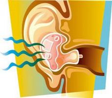
ototoxicity picture

Ototoxicity Symptoms, Signs

Ototoxicity signs and symptoms in adults include problems in hearing and it is usually bilateral or coming from both ears. Then, they may also feel as if something is ringing in their ears or tinnitus. At the same time, they will also sense a loss of balance making it difficult for them to walk in a straight path.

For kids however, they may not talk properly so parents wouldn’t know what the problem is especially if they are under medications. Parents should take important cues if their kids have hearing problems like having difficulty hearing whenever their names are called. Sometimes it also affects the speech of the child since he or she is not able to hear things properly. As a result, they find it difficult to talk. Also, they become inattentive and have problems with learning especially when they are sent to school. Also, a noticeable sign from children with ototoxicity is that they turn the volume louder when listening to music or watching television. They also don’t respond nor turn their heads when hearing a sudden loud voice like banging of the door or utensils falling on the ground.

To some extent, in worse cases, even with a simple standing up position or walking, the patient feels “woozy”. They also feel a sudden blurring vision with a simple move of their head called as oscillopsia. They will also feel nausea and vomiting, dizzy, syncope and even elevated blood pressure.

Ototoxicity Causes and Risk Factors

Ototoxicity is almost always caused by drug reactions. There are different types of drugs that cause hearing problems and imbalance to persons who are taking this. What is quite alarming is that even over the counter medications can cause ototoxicity so patients should be advised to always read the drug interactions and possible side effects to avoid this problem.

1. Aminoglycosides ototoxicity

Aminoglycosides is a class of antibiotics that can cause ototoxicity.  Usually these drugs are given via intravenous route so there is a greater chance that it can cause ear damage since the drug passes through the blood stream. Some of the popular drugs prescribed by doctors include gentamicin, streptomycin, neomycin and others.

2. Vancomycin ototoxicity

Vancomycin is an antibiotic that is mostly used as a prophylaxis for treating gram negative bacteria. usually, this drug is the “last resort” when all other antibiotic are used but have failed to treat the patient from the infection. For patients with pseudominas infection, if metronidazole is not effective, this potent drug is prescribed by doctors.

3. Cisplatin ototoxicity

Cisplatin is a chemotherapeutic or anti-neoplastic drug that is used to treat cancer. Amongst the cancer drugs, cisplatin is very famous to cause ototoxicity. The good news is that this drug is dose-dependent. If the dose is higher than 60 mg, the greater risk for having ototoxicity through cisplatin.

4. Furosemide (Lasix/Loop diuretic)

Furosemide is a known diuretic wherein it is used to help excrete the excess water in the body. Furosemid is ototoxic when the treatment is suddenly stopped or when the dosage of the drug is more than 240mg.

5. Aspirin ototoxicity

Aspirin along with other quinine drugs can cause ototoxicity where patients will be able to experience hearing problems either bilateral or unilateral.  Also, with aspirin, patients will also experience ringing of the ears or tinnitus

6. Azithromycin ototoxicity

Azithromycin is another antibiotic which is a type of macrolides and this drug is famous to treat sore throat. This can cause ototoxicity but the effects can be reversed if the patient stopped taking the drug.

7. Vestibular ototoxicity

With vestibular ototoxicity, the problem is mostly on the sense of balance because the vestibular system is widely affected with the effects of medication

8. Methotrexate ototoxicity

This is also another is mostly used to treat cancer like cancer of the lungs, skin, breast etc. and this drug is also known to have serious side effects like ototoxicity.

Ototoxicity Diagnosis

When patients consult the doctor for ototoxicity, without having to take blood samples and others tests, they can likely diagnose ototoxicity judging from the medications taken.

Audiogram

In order to make sure and detect the severity of the problem, sometimes audiogram is conducted by ear specialist or ENT doctors

With audiogram, the patient will be given an audiometer where they will hear different frequencies via the earphones. The range of the frequency is from 100Hz to 8000Hz. Then the result is plotted and measured and the doctors will interpret the result.

Ototoxicity Treatment

Withdrawal of drug

Sadly, there is no definite treatment for ototoxicity yet. Although withdrawal of the drug may reverse the effect but there are no scientific proofs yet. As of today, the treatment goals are more on stopping or reducing the harmful effect of the drug to avoid total hearing loss and imbalance problems.

Preserve hearing

  1. Hearing aids
  2. cochlear implants

The above are suggested treatments to help individual preserve their sense of hearing.

Physical therapy

For ototoxicity people with balance problems, physical therapy is also advised to help them and their brain become accustomed on the ways to maintain balance when standing up or walking around.

No responses yet

« Prev - Next »

© 2017 eHealthWall.com. All Rights Reserved. Privacy Policy
This website is for informational purposes only and Is not a substitute for medical advice, diagnosis or treatment.