Archive for April, 2012

Primary Peritoneal Cancer – Symptoms, Prognosis, Survival Rate, Life Expectancy

Apr 21 2012 Published by under Diseases & Conditions

What is Peritoneal Cancer?

These days, cancers have become aggressive and the symptoms are vague making it very difficult to treat in the early stages. One of it is the peritoneal cancer. This is one of the rarest forms of cancer wherein it affects the peritoneum. The peritoneum is a thin sheet which is found in the walls of the whole abdomen and it even extends and covers the rectum and bladder as well. The peritoneum’s main function is to help the digestive tract move in a smooth manner through the lubricating fluid it secretes.

Peritoneal cancer occurs primarily in women. In fact peritoneal cancer mimics the same signs and symptoms with ovarian cancer. This is because there are genes that link the mutation of ovarian and peritoneal cancer.

Peritoneal cancer

Primary Peritoneal Cancer

This type of cancer is one of the rarest of its own. It is a cancer of the cellular lining located in the peritoneum, or what is called as the cavity of the abdomen. It covers both the uterus and the abdomen. It also extends over the uterus and bladder. The peritoneum is known as a protector of the abdominal contents and it also a producer of fluid for lubrication. This fluid aids in the abdominal organ to smoothly move within the abdomen as we go on our day to day activities.

It occurs particularly anywhere within the abdominal area and affects the any organ which can be located in that vicinity. Women who are diagnosed with this disease condition, treatment and symptoms manifested by these women are the same with those women having ovarian cancer.

Secondary Peritoneal Cancer

With regards to this type of cancer, its etiology is due to the fact that the cancer, itself, has spread into various areas of the body. Diagnosed with this kind of disease will give you a lesser rate of survival. It has a lower survival rate because it means to say that the cancer was not spotted earlier. Hence, it progressed to a secondary state.

With regards to the treatment, it will follow the basic three step treatment for cancer, which is chemotherapy, surgery, and radiation. If the cancer can still be managed with surgery, surgery is done plus chemotherapy. On the contrary, if the cancer has already spread and can’t be managed anymore by surgical procedure, the surgeon or the physician will then suggest that the patient undergoes radiation therapies with chemotherapy.

Peritoneal Cancer Symptoms

Generally, the symptoms exhibited by peritoneal cancer seem general in the beginning. A lot of doctor suspect the patients with ovarian cancer since the signs and symptoms are the same. But with peritoneal cancer, the symptoms include:

  • Loss of appetite
  • Feeling very full after a light meal
  • Nausea
  • Diarrhea
  • Abdominal pain or discomfort wherein the patient will feel cramps, bloating and sometimes indigestion and gas
  • Weight gain or weight loss
  • Vaginal bleeding
  • Ascitis or accumulation of fluid in the abdomen

Peritoneal Cancer Diagnostic Tests

The symptom ascitis is commonly reported amongst the symptoms that were presented earlier. When ascitis occurs, it is when the patients would seek help for peritoneal cancer is too late. Hence, upon the consultation with a physician, it will be too late to know that the disease itself has already spread elsewhere. Aside from the usual physical exam, ultrasound and blood works, CA-125 Assay is the tumor marker for peritoneal cancer. Along with that are the other diagnostic test that will help in diagnosing this disease condition are as follows:

  • Pelvic Exam – With this kind of diagnosis, the physician will inspect through palpating the different organs to be able to find unusual shapes or sizes.
  • Ultrasound – This test is done with the use of high frequency waves of sound. This is aimed at the ovaries. The echoes produced in this type of diagnostic test will create a picture that will give a view of the organs inside the abdomen; such picture is called as sonogram.
  • CA-125 Assay – This is an important tumor marker that is found in the blood. When this marker is present, it is used to diagnose a positive cancer in both ovarian and peritoneal.
  • CT scan – What happens here is that there is a series of précised pictures of the parts inside the body. It is done through the aid of a computer that is connected to an x-ray machine.
  • Lower GI series or Barium Enema – After giving the patient with enema that is a chalky white solution that has barium, the pictures are then taken through series of x-rays of the colon and rectum. The solution used here outlines the rectum and colon on the x-ray which will make it easier to spot any abnormal areas.
  • Biopsy – This particular kind of test is done through getting a sample from the suspected area that is to be examined under microscopic equipment, which is usually studied by a pathologist. The test will be slightly painful. Needle biopsy is usually performed by some physician. In order to get a sample tissue, the surgeon can perform laparotomy, wherein the surgeon opens the patient’s abdomen. If suspected with cancer, the surgeon may remove the cancerous organ to prevent it from spreading.

Peritoneal Cancer Treatment

Once peritoneal cancer is diagnosed, the treatment depends on different factors like:

  • The patient’s age and health condition
  • The location of cancer and the size
  • Cancer grade and stage

When cancer is detected earlier, surgery and chemotherapy may be done. But this also depends on the overall condition of the patient. With chemotherapy, it is crucial that the patient can withstand the painful chemotherapeutic drugs. The chemo drugs not only destroy the cancer cells but the normal cells as well. This results to compromised immunity making the patient susceptible in infections. The drugs used in treating peritoneal cancer are also the same drugs treating ovarian cancer. Sometimes, the drugs are delivered directly into the abdominal area to directly kill cancer cells.

Surgery is also another option for treating peritoneal cancer. If the tumors are visible and it does not affect the other organs, surgery may be done. But at some point, women’s reproductive organs are also removed such as the uterus, ovaries and the fallopian tube.

But if the peritoneal cancer is discovered at the later stage of the disease, doctors suggest doing palliative treatment to help prolong and preserve the patient’s quality of life. The health care teams assists in giving comfort from the cancer pain, managing weight loss and even addressing the fluid accumulation due to ascitis.

With regards to the staging of cancer, there are actually four stages. Stages simply mean the size of the cancer itself, whether or not it is spreading throughout the body.

Stage 1 is when a small area is affected and the cancer cells have not yet spread. Meanwhile patients diagnose as having Stage 2 of this cancer, means that the cancer has already spread, and is beginning to grow. However, it may or may not be detectable. The peritoneal cancer is usually not early detected. Hence, when discovered, the patient will be either in stage 3 or stage 4. In stage3, the tumor is already present. It is located within the abdominal cavity. However, when one reaches stage 4, it becomes more dangerous. This is when the cancer has already metastasized or spread to other organs which are located outside the abdomen. Such organs like liver or lungs and the like.

On the other hand, the term grade is given to patients by their physician due to the fact that it will describe the growth of the cancer cells. There are 3 grades. Grade 1 is when the growth of cancer cell is slow. Under a microscope, it mimics the normal tissue. Grade 2, on the other hand, looks also like normal cells. However, it grows and spreads in a rapid manner. The last grade, grade 3, is sometimes classified as the aggressive cancer cell. It looks bizarre, grows and spread very fast.

Prognosis

The one thing that you have to take note is when a person is diagnosed earlier with peritoneal cancer, the better the prognosis for that person. Meanwhile, people diagnosed with secondary peritoneal cancer have a poor prognosis to begin with. The prognosis for this kind of cancer will focus on the ability to treat the primary cancer. If given the chance that the primary cancer can’t be surgically removed or treated, the physician should get the remaining cancerous cells in order for the improvement of health of the patient and for the optimum good of the patient’s general condition.

Survival Rate

In comparison to other type of cancers, the peritoneal cancer survival rate is very low. However, with the high technology and the various cancer centers all over the world, the survival rate for these patients has increased. This kind of improvement is a good sign.

With regards to the peritoneal cancer survival rate in stages 3 and 4, both are almost the same. They have progressively slower results. Yet with the advance studies, the rates for the survival of these patients have a possibility to become better. The right treatment is the primary key to it.

The survival rate for stage 3 is low. It is in this stage where the cancer has already spread. Despite of that information, the patient can still increase the chance of survival if the body is accepting the treatment which leads to results that are good.

Just like stage 3, stage 4 rate of survival are very low. Studies show that few patients had been able to reach this stage. This is considered to be the most advanced cancer stage. It is known as Duke’s D colon cancer or metastatic cancer. The survivors of this stage cling to persistent and very effective treatment.

Life Expectancy

The life expectancy for this kind of cancer primarily boils down to the stage of the disease, the age of the patient, the general health condition of the patient.

In general, some would say that the median average life expectancy of a patient having this kind of cancer will ranges between 1 year to 2 years and a month. In extreme cases, it can range from 4 months to 5 years.

No responses yet

Pregnancy Symptoms Week by Week

Apr 20 2012 Published by under Pregnancy

Pregnancy symptoms week by week is one of the concerns of pregnant women because they want to know what is happening to their bodies as well as what is happening inside their womb. This article discusses the pregnancy symptoms week by week as well as the fetal development week by week. It shows the possible pregnancy symptoms week by week in order for expecting mothers to expect and prepare for the changes throughout pregnancy. The most important thing of knowing the changes throughout pregnancy is to maintain a healthy one with good pregnancy outcomes.

Pregnancy Symptoms week by week

Week 1

Maternal Changes – During this week, the body is getting ready for ovulation.

Fetal Development – There is still no fetus that is developing, but the ovaries are starting to develop a mature egg cell for ovulation.

Recommendations: Women planning to have pregnancy should start taking iron and folic acid supplements as soon as the last menstruation has ended in order for the body to prepare for possible pregnancy during the next cycle. It’s also nice to know the exact date of ovulation through menstrual calendars in order to determine the right time to conceive.

Week 2

Maternal Changes – The uterus has just developed the uterine lining in time that the egg cell will be fertilized by a sperm. During the time of ovulation, women may feel a sharp pain on side of the lower abdomen as a sign of egg cell rerelease known as mittleschmerz as a sign of pregnancy symptom week by week.

Fetal Development – The ovum is now released in the fallopian waiting for a sperm cell for fertilization.

Week 3

Maternal changes – Fertilization has just occurred after a successful sexual intercourse. There may be signs of implantation because the zygote or the fertilized egg implants at the lining of the uterus. Spotting during this time should not be mistaken as menstruation. Mothers should abstain from drinking alcohol and smoking because these may have effects on the growing embryo. Smoking during pregnancy may bring about problems in fetal development. The mother’s body also releases early pregnancy factor to prevent the body from attacking the embryo as a foreign body.

Fetal Development – The zygote divides rapidly as it travels along the fallopian tube into the uterus for implantation. The zygote usually travels to the uterus for 4 to 7 days. Cell division occurs until it becomes a blastocyst. On day 10, the blastocyst has successfully implanted. Once implantation has occurred, the zygote is now known as an embryo.

Week 4

Maternal Changes – During this time, a pregnancy test will usually yield a positive result due to increase in a hormone called HCG or Human Chorionic Gonadotropin. Women may also start feeling presumptive signs of pregnancy such as mood swings, frequent urination, tender breasts and feelings of tiredness.

Fetal Development – The first trimester is the most crucial stage for the fetus because organogenesis happens or the development of the organs. The 4th week of pregnancy reveals a yolk sac that acts as a placenta.

Week 5

Maternal Changes – Pregnant women may start feeling headaches, crampy uterus and morning sickness. This usually persists until the end of the first trimester.

Fetal Development – The heart of the bay begins to beat early during this week. This may be apparent in ultrasounds. The head and the tail of the baby are also distinguishable. The neural folds also begin to fuse to form the spine. The average length of the fetus is now 1.5 to 2.5 mm.

Week 6

Maternal Changes – The morning sickness may get intensified during this week because of continuous rise is maternal hormones. The areola will also start to appear darker and bigger. Avoid cleaning the cat litter to prevent toxoplasmosis that can result in fetal abnormalities.

Fetal Development – The larynx and the inner ear start to appear. The limbs also develop as buds. The heart begins to bulge and the circulation is well established. The placental lining is still forming. The primordial of the stomach, lungs, pancreas and liver may also be evident.

Week 7

Maternal Changes – Chloasma or melasma start to appear because of increasing pregnancy hormones.

Fetal Development – The baby is about 7 to 9 mm during this stage. The hand plates are present and the genital tubercle is also developing, but it is still hard to distinguish if the fetus is a boy or a girl.

Week 8

Maternal Changes – Regular prenatal check-ups start at this week. The maternal symptom such as morning sickness, feeling of tiredness and breast tenderness still persist.

Fetal Development – The length of the baby is 8 to 11 mm from crown to rump. The hind brain of the fetus is visible, the gonads will differentiate into either ovaries or testes and there is spontaneous movement, but the mother may still not feel it. Hardening of the bones also starts during this stage and elbows start to appear. The toes may also have developed by now.

Week 9

Maternal Changes – Mothers may begin feeling stuffy nose because of increased progesterone in the body. Nose bleeding and nasal congestion may be normal during this stage.

Fetal Development – The fetus already has distinct elbows by now. The length will be about 13 to 17 mm. The average weight will be also 1 gram. The gonads of the fetus have already developed.

Week 10

Maternal Changes – The abdomen may begin to enlarge a little as a pregnancy symptom week by week during this time. The waistline will slowly disappear and linea nigra, a black, thin line from the umbilicus to the symphysis pubis, may develop. This integumentary change may eventual disappear weeks after delivery.

Fetal Development – The embryo now becomes a fetus during the 10th week of pregnancy. It has grown to 25 to 35 mm in size and increases weight to 4 grams. There are already evident tiny toes. The eyes may be largely open; however, the eyelids may still begin to fuse. The genitals of the fetus may also start to differentiate, but still not distinguishable. The upper lip and the external ears also have formed. The tail of the fetus already has disappeared during this stage.

Week 11

Maternal Changes – Gaining weight starts to begin during this stage. The normal weight gain for pregnant women is 1 to 2 lbs a month. Healthy diet is a key to prevent excessive weight gaining.

Fetal Development – The head of the baby is actually 3 times larger than the body. The fingernails may appear during this week and the iris of the eyes is developing. The average weight of the fetus during this week is 7 grams.

Week 12

Maternal Changes – The early symptoms of pregnancy such as nausea and extreme tiredness usually fade away as the first trimester ends. The abdomen will also start to appear bigger. The placenta has been fully developed and it takes the place of the corpus luteum in producing maternal hormones. Risk for miscarriages is reduced at the end of the third trimester.

Fetal Development – The structure of the brain is now similar to the brain at birth. The liver starts to secrete bile during this week. The average weight doubles from the previous week to 14 grams. The length will now be 3.5 inches. The reflexes also appear. Doppler may be able to detect the heart beat of the fetus.

Week 13

Maternal Changes – This is the beginning of the second trimester. During this time, the mother feels stronger because of absence of morning sickness and other uncomfortable feelings. Pregnancy starts to become real for spouses.

Fetal Development – The complete set of baby teeth has developed, but still has not erupted until the 6th month of extrauterine life. The average weight is 28.3 grams. The placenta also continues to grow. The intestines begin to migrate into the abdominal cavity. Insulin has also stared to be secreted by the pancreas. The presence of diabetes in mothers may be detected by the pancreas of the fetus, which secretes more to cope with the maternal blood. The fetus’ stool or meconium also begins to develop on the intestines.

Week 14

Maternal Changes – Chloasma is starting to be more apparent during this time as estrogen and progesterone continues to increase. The linea nigra may be darker and more apparent as well as the areola also starts to become bigger.

Fetal Development – The size of the fetus is now 12.5 cm. The kidneys are actually producing urine. The urine may mix with the amniotic fluid. The fetus starts to ingest the amniotic fluid and eventually excretes it through the urine.

Week 15

Maternal Changes – The heart increases cardiac output to compensate with low hemoglobin levels and to supply the baby with blood through the placenta. There is also increasing weight and the usual clothes may not fit well anymore. Detecting fetal abnormalities through blood tests such as Alpha-fetoprotein determination can now be done.

Fetal Development – The skin of the fetus is very thin with visible blood vessels. The average weight is about 70 grams. The heart begins to pump more blood of about 25 quarts of blood in a day. The hair pattern on the scalp also develops.

pregnancy symptoms week by week image

Pregnancy symptoms week by week picture
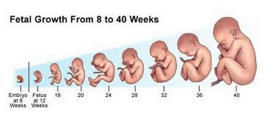
fetal development week by week from 8 to 40

Fetal development week by week

Week 16

Maternal Changes – Women may have increased sexual drives because of very high maternal hormones. Modifications in positions may be necessary, but generally, sexual intercourse is not prohibited during pregnancy.

Fetal Development – The eyelashes and the eyesight of the fetus have already developed during this stage and the nails in the fingers and toes have started to grow.

Week 17

Maternal Changes – The fundus of the uterus is now between the navel and the symphysis pubis. The vaginal secretions, sweating and nasal congestion may increase normally.

Fetal Development – Brown fats are starting to develop at the skin. Brown fats help fetus maintain normal body temperature when they are born. The average weight is now 142 grams. Startling reflex may be apparent when the fetus hears loud noises on the outside world.

Week 18

Maternal Changes – Since the uterus may compress other organs when you lie down, women now start to sleep harder. Sleeping with more than one pillow usually relieve this feeling. Increased urination is also more intense as the growing uterus pushes into the bladder.

Fetal Development – The fetus is growing rapidly during this stage. The bones still continue to harden. The fingerprints may be well developed and the pads of the toes and pads are developed.

Week 19
Maternal Changes
Heart burn and indigestion may be a special problem starting this week because of slowed intestinal motility and gastric emptying as a result of maternal hormones. Staying upright after eating is a remedy to prevent heart burn.
Fetal Development
The ovaries of female fetuses normally may have developed primitive egg cells during this time. Fine hair called lanugo also grows in the skin of the fetus. Lanugo normally lessens as the baby reaches term.

Week 20

Maternal Changes – This stage may make mothers excited because they are halfway over pregnancy. The navel may start to prop out because the growing uterus pushes out the belly button. There is also increasing difficulty of breathing as the uterus pushes against the diaphragm. During this time, the fetus may have quickening, meaning that it moves constantly inside the womb making the mother notice the movement. Mothers usually feel becoming a mother for the first time when life begins to be apparent inside the womb.

Fetal Development – A creamy, cheese-like substance on the skin called vernix caseosa begins to form. Vernix is more apparent in the creases in the body. This protects the baby from cold temperature at birth. The weight of the fetus may now be 283 grams with a length of 25 cm. The 20th week may be the best time to have an ultrasound to look at the gender of the baby.

Week 21

Maternal Changes – Stretch marks may start to appear as the skin starts to stretch to make way for the growing abdomen. Back pain also intensifies because of increased weight of the fetus and placental structures.

Fetal Development – There is no new formation during this stage. The fetus may weigh 369 grams.

Week 22

Maternal Changes – Libido may still be increased during this time because of increased blood flow into the clitoris and the vagina.

Fetal Development – The fetus continues to develop the eyebrows. The average size during this time is 27.5 cm with a weight of 425 grams.

Week 23

Maternal Changes – Swelling of the feet may be apparent because of pressure of the uterus to the major veins on the lower extremities. This may be normal, but swelling in the face and hands may mean more serious indications. Women may also feel slight discomforts from Braxton Hicks contractions or faint contraction of the uterus in preparation for childbirth.

Fetal Development – The lanugo usually darkens. The weight increases to 1 pound.

Week 24

Maternal Changes – The fundus of the uterus usually is in line just above the navel with increasing difficulty of breathing. During the second trimester, there may be lesser frequency of urination than the first trimester because the uterus usually ascends to the pelvic and abdominal cavity.

Fetal Development – Brown fat still continues to be deposited under the skin. The weight of the fetus may be 596 grams or 1.5 pounds. The fetus also grows to up to 12 inches in length.

Week 25

Maternal Changes – This is the beginning of the third trimester. At this point there is increased weight gaining of up to 1 pound per week because of continuous growth of the fetus.

Fetal Development – The bones still continue the hardening process or ossification. The fetus may weigh slightly heavier of 709 grams.

Week 26

Maternal Changes – Sleeping may be especially difficult for women because of the compression of the vena cava. Pregnant women are advised to sleep on their left side to prevent dizziness and hypotension as a result of compressing the vena cava.

Fetal Development – The fetus may start to hear the surroundings on the outside world. In this line, listening to music may help stimulate the baby. The fetus may also be able to see light and dark as light may pass through the skin into the uterus.

Week 27

Maternal Changes – The breast can weigh as much as 14 ounces because of engorgement in preparation for lactation. The amniotic fluid will also increase in amount as evidenced by increased weight.

Fetal Development – The skin of the fetus is usually wrinkled due to presence in amniotic fluid all the time. The wrinkles usually disappear days or weeks after birth. The baby may be as heavy as 900 grams.

Week 28

Maternal Changes – Increase prenatal check-ups are required to at least every two weeks. The breast may also start to produce colostrums, essential breast milk for babies that contain antibodies.

Fetal Development – Subcutaneous fats start to be deposited. Descent of testes among boys may also begin. The baby may weigh up to 1 kilogram and grow as large as 35 cm.

Week 29

Maternal Changes – Increasing discomforts on the back, legs and breathing is starting to get intensified. It is essential to get proper exercise, rest, nutrition as well as good posture to prevent musculoskeletal problems such as cramps, and back aches.

Fetal Development – The bone marrow is fully developed to produce the baby’s own red blood cells and other blood components. The kidneys may also produce up to half a liter of urine everyday.

Week 30

Maternal Changes – Same feelings may affect the mother such as back aches and leg pains. Exercise and good posture is a key to reduced symptoms.

Fetal Development – During this time, the fetus may grow up to 1.36 kilograms and may have length of 37.5 cm.

Week 31

Maternal Changes – The rib cage may be sore because of pressure of the growing fetus in the rib cage. The fetus may also fill up the space in the abdominal cavity displacing all organs inside.

Fetal Development – The iris and pupils of the fetus may now react to light. The skin of the fetus is usually pink. It now weighs up to 1.5 kg. The fingernails may also reach up to the end of the fingers or toes.

Week 32

Maternal Changes – Mothers may be especially concerned about labor as pregnancy comes to end. It is important to discuss fears and concerns to other people as well as to the doctor.

Fetal Development – There may be an increased amount of hair in the scalp. The average weight of the fetus may be 1.7 kg with a length of 16 inches.

Week 33

Maternal Changes – Small bumps may be felt on the abdomen while the baby moves inside the uterus.

Fetal Development – The lungs have already formed surfactant, which helps the baby breathe after birth. When a baby will be born during this stage, the chances of survival are higher than earlier weeks because of the presence of lung surfactant.

Week 34

Maternal Changes – There may be more frequent Braxton Hicks contractions, which help women, prepare for birth. There may be breast milk production as the hormones in the placenta stimulate breast milk production.

Fetal Development – The baby may reach 2 kg in weight and 16.8 inches in length. The baby may also urinate more in the amniotic fluid.

Week 35

Maternal Changes – Increase difficulty of breathing is felt during this time. There may be increased urination, but less often than what happens as you reach term.

Fetal Development – The baby may weigh at least 2.15 kg getting ready for birth. The length may reach 18 inches. The organs have been developed during the first trimester, but finishing touches occur during this time. The baby’s brain may also begin to double in size.

Week 36

Maternal Changes – Prenatal check-ups should be increased to once a week in order to prepare for childbirth. The mother may be relieved of difficulty of breathing as the fetus descends down the pelvis. However, increased urinary frequency is more intense as the fetus pushes more on the bladder.

Fetal Development – The baby may assume a head down position compatible for vaginal delivery. During this time, the fetus also descends into the pelvis ready for delivery. The baby may weigh 2.3 kilograms and has a length of 18 inches.

Week 37

Maternal Changes – The pregnant woman is considered to have reached term staring the 37th week of pregnancy. It is important to prepare the things for birth because the fetus is ready for birth anytime.

Fetal Development – The baby practices breathing by inhaling amniotic fluid in the lungs.

Week 38

Maternal Changes – The woman may feel tingling sensations in the vagina and the legs as the baby’s head settles into the pelvis for delivery as pregnancy symptoms week by week during this stage.

Fetal Development – The weight gain for the fetus during this time is one ounce in a day. There is a large growth happening in the fetus during this time.

Week 39

Maternal Changes – One week left and you’re going to give birth. Some women may have given birth at this time and it is considered term and safe. Lightening may be experienced as the fetus descends further into the true pelvis. Expecting others may present symptoms during this week or few hours prior to labor such as:

  • Bloody show in the vagina as a result of loss of mucous plug
  • Loose stools
  • Increase in appetite
  • Sudden increase in energy

Fetal Development – There are no developments happening on the fetus during this time because of imminent delivery. The meconium may reach the rectum ready to be released at birth or few hours after birth.

Week 40

Maternal Changes – The mother is now ready to give birth. The actual percentage of women giving birth on their due date is only 4%. Most of women give birth 2 weeks before or two weeks after expected date of delivery.

Fetal Development – Most of babies during this week have assumed a cephalic presentation for vaginal delivery. However, up to 4% may assume different presentations such as breech or shoulder that may need cesarean birth to prevent fetal distress.

These detailed pregnancy symptoms week by week provides knowledge to pregnant women. These pregnancy symptoms week by week should be observed in order to detect any abnormalities in the mother as well as the fetus. The pregnancy symptoms week by week should also be reported to the physician in order to provide managements for the discomforts.
No responses yet

Occipital Neuralgia – Symptoms, Treatment, Causes and Surgery

Apr 20 2012 Published by under Diseases & Conditions

What is Occipital Neuralgia?

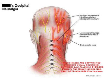
It is otherwise known as Arnold’s neuralgia or C2 Neuralgia. It is a medical term given that connotes a cycle of having pain then spasm then pain again which originates from the base of the skull or medically termed as suboccipital area which radiate to the person’s back then to the front and towards the head’s side and also behind the person’s eyes.

It is a condition that affects the neurological aspect in which the ones affected are the occipital nerves which are injured or inflamed. It is similar to migraine or headache because they produce similar symptoms which can confuse you. The main difference is that this condition is a distinct kind of disorder that needs a proper treatment.

Other experts would define it as a peripheral nerve stimulator or a headache form involving the occipital nerves which emerges between the spinal bones located in the upper neck region towards the scalp area. It is often characterized as having a pain paroxysm that occurs with the distribution of the person’s occipital nerve.

Occipital Neuralgia

Occipital Neuralgia image

Occipital Neuralgia Symptoms

One particular symptom associated with this condition is having a chronic form of headache. Another notable symptom is the presence of intense like pain which is often described as jabbing, electric shock, sharp pain in the person’s back of the neck and head. Other symptoms that the patient may experience include:

  • Light sensitivity
  • Scalp tenderness
  • Pain behind the person’s eye
  • Pain when moving the person’s neck
  • Chronic form of HeadacheIntense headache Image

Chronic form of headache (symptom of occipital neuralgia)

  • Pain upon moving the neck
  • Pain on both or one side of the person’s head

These are just the common symptoms associated with this disease condition.

Occipital Neuralgia Causes

The occipital neuralgia may either be classified as primary or secondary. When a person has a primary kind of occipital neuralgia, they don’t have a known cause. Since there is a no known cause or it is idiopathic in nature, it is often confused with cluster headaches or migraine. On the other hand, when a person has secondary kind of occipital neuralgia, they have the disease because it is associated with a disease condition such as:

  • Tumor
  • Infections
  • Hemorrhage
  • Trauma
  • Repetitious contraction of the neck
  • Nerve stress
  • Nerve lesion
  • Arthritis
  • Always keeping the head forward or downward position in a lengthy period
  • Gout
  • Diabetes
  • Inflammation of the blood vessel
  • Contraction of the muscle neck which is repeated
  • Compression of the spinal column

These are the common causes of why persons acquire the disease condition called occipital neuralgia.

Occipital Neuralgia Treatment

The treatment for persons who have occipital neuralgia will depend on the cause of why the person has the condition. The primary goal is the reduction of inflammation and the relief of pain. You can relieve your pain through:

Non Surgical Management

  • Resting in a quiet room – Having a massage especially in the painful and tight muscles of the neck
  • Over the counter anti inflammatory medications such as ibuprofen
  • Applying heat to the person’s neck
  • Acupuncture – The care modalities mentioned above are the one that is tried out first before going through the physician drug prescription.

Medical Treatment

If the independent care modalities do not work, your physician may suggest or prescribe the following treatment drugs such as:

  • Antidepressant drugs which is considered as the main and primary treatment in preventing may types of headaches which includes occipital neuralgia.
  • Steroid drugs which is for short term use only which is used for inflammatory reduction
  • Anti neuropathic drugs to prevent and treat neuropathic problems
  • Anticonvulsant medications such as Neurontin and Tegretol to prevent convulsion
  • Muscle relaxants that are under the prescription drugs given to relax the tense muscles
  • Anti inflammatory medications such as non-steroidal anti inflammatory drugs given to reduce the inflammatory process
  • Analgesic medication given to treat pain
  • Occipital or Local nerve block which involves in the injection of a medication which numbs the pain

Surgical Management

  • Peripheral nerve stimulation to stimulate the nerves
  • Occipital Neuralgia Surgery – When, for instance, the care modalities or treatment provided, as stated above, does not work as it should and as it is expected to be. Then the last option is surgical procedure which may include either of the following:

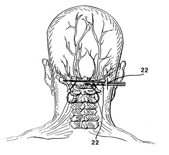
Occipital Nerve Stimulation –  What happens here is that, there is a usage of neurostimulator which delivers the electrical impulses towards the person’s inflamed or destroyed occipital nerves. Here, the insulated wires are being attached to the neurostimulator which are being inserted under the person’s skin. Through the electrical impulse, it can aid in the blocking of the pain message which is brought through the person’s brain. This kind of surgical procedure is a less invasive kind which will not damage the areas surrounding the nerves and the nerves itself.

Peripheral Nerve Stimulation Image

Occipital Nerve Stimulation

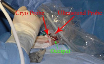
Decompression of the Microvascular Nerve – What happens here is that the doctor or the surgeon in charge relieves the pain through adjusting the person’s blood vessels which compresses the person’s nerve. Here there is an exposure of the occipital nerves and the surrounding vessels. What is done is that the vessels are being moved away from the person’s nerves which will lead to less pressure on the nerves leading to a better functioning of the nerves. This kind of surgical procedure acts like a pacemaker of the heart, which functions as a deliverer of small charges of electricity to the patient’s nerve in order to prevent the occurrence of any type of headache. In other case, the surgical procedure of burning the nerve is done instead of cutting it. The main goal of surgical procedure is to cut the nerve in the occipital region which will prevent it to send messages especially pain messages to the person’s brain. Before undergoing any of the procedures mentioned, the patient and the physician should be able to weigh the pros and cons or the benefits and risk before deciding to pursue the surgical procedure.

surgical procedure image

Surgical procedure to treat Occipital Neuralgia

No responses yet

Dyscalculia – Definition, Symptoms, Test and Treatment

Apr 19 2012 Published by under Diseases & Conditions

What is Dyscalculia?

Dyscalculia in children and adults is a learning disability that results in problems with arithmetic. Dyscalculia definition means that a person with dyscalculia learning disability has poor mathematical skills. Dyscalculia is genetic in cause or could be acquired. It may also be developmental as the child learns how to deal with numbers.

Dyscalculia learning disability involves difficulty in manipulating numbers, understanding them and learning arithmetic such as simple addition or subtraction.

The learning disability is not confined to people with low intellectual abilities. In fact, cases occur in a wide range of IQ levels. Problems encountered by people with dyscalculia also include measurement, time, and spatial skills. Dyscalculia also involves both problems on abstract reasoning, calculation and memory with numbers.

Dyscalculia Symptoms in children

Source – Buzzle.com

Dyscalculia Signs and Symptoms

Dyscalculia presents early as a problem in subtizing. Subtizing is the capacity to identify how many objects are there in a small group, without even counting them. This aptitude in knowing how many objects are there at a glance is already developed at birth. Infants can already subtize up to three objects. People with dyscalculia find it hard to subtize or takes time to subtize as compared to normal people of same age.

Other signs and symptoms include:

  • Problems in identifying time using analogue clocks
  • Problems in arithmetic including difficulties in addition, subtraction, division or multiplication tables
  • Adults find it hard to understand budgeting and financial planning
  • Difficulty in identifying left and right
  • Exceptional in writing. Most people with dyscalculia has excellent writing skills.
  • Difficulty in understanding maps
  • Problems in estimating distance
  • Over sensitivity to external stimuli
  • Difficulty in handling money and identifying change
  • Problems on sequencing and understanding value
  • Inability to decipher concepts of weeks, days, months, quarters and seasons
  • Children may find it difficult to line numbers

Causes of Dyscalculia

Several factors are being considered causative factors of dyscalculia learning disability. These include:

  • Problems on working memory – Faults in the working memory contribute to the occurrence of arithmetic problems.
  • Neurological problems – Injury to the junction of the parietal and temporal lobe also may lead to dyscalculia. Injury includes presence of lesions in the area or through mechanical blows.
  • Heredity – Genetics have also played a role in the occurrence of dyscalculia.
  • Disturbance in short-term memory – This makes the person unable to remember arithmetic calculations or formulas.

Types of Dyscalculia

Dyscalculia learning disability presents in various types such as:

Lexical Dyscalculia

Lexical dyscalculia learning disability involves the ability to understand mathematical and arithmetic ideas during articulation, but unable to understand them when they present in equations or problems. Individuals with dyscalculia can read numbers, but unable to remember them in a larger picture.

Verbal Dyscalculia

Verbal dyscalculia involves the inability to articulate mathematical ideas or concepts. The person has no problems with writing and reading numbers and equations.

Graphical Dyscalculia

Graphical dyscalculia involves the inability to write numbers, equations or symbols. The person understands arithmetic, but unable to write them to express understanding.

Practognostic Dyscalculia

Practognostic dyscalculia involves the inability to use mathematical concepts in practical applications such as in day to day living. The person can read, write and articulate the concept, but cannot apply it.

Ideognostic Dyscalculia

Ideognostic dyscalculia is the general inability to understand math in a holistic manner.

Operational Dyscalculia

Operational dyscalculia is the inability to perform mathematical calculations such as addition, subtraction, division or multiplication. The person understands the numbers, but cannot manipulate them in operations or calculations.

Diagnosis and Assessment for Dyscalculia

Diagnosis of dyscalculia involves several tests to determine the presence of the learning disability. Dyscalculia test often require a paper and pencil test. Further tests are required to reveal how a person practically applies mathematical concepts. Results are compared with the expected skill based on educational level and age.

Areas that are assessed during dyscalculia tests include:

  • Skills in adding, subtracting, dividing, multiplying and counting
  • Skills in identifying when to use mathematical operations
  • Skills in organizing objects
  • Skills in measuring time, money, volume, and other quantities
  • Skills in re-checking own work and using alternatives to come up with a correct solution

Dyscalculia screener devised for teachers is also available to let teachers identify dyscalculia among children. This screening tool identifies dyscalculia outside other areas of learning such as language, reading and writing.

A dyscalculia online test is also available to initially assess presence of dyscalculia learning disability. However, it is only a screening tool for children who might have the condition. Further assessments should still be done my professionals.

Dyscalculia Treatment

Treatments for dyscalculia learning disability include the following measures:

1. Educational Therapy

Conventional management for dyscalculia aims at improving the skills of the child in terms of arithmetic and math. Trained teachers or special education teachers usually conduct this type of therapy.

Educational therapy involves modification of teaching approaches to children. One of these is the use of graphing paper to help children present concepts in an organized manner. Simple memorization of the multiplication table may also be modified by explaining the meaning of the numbers.

2. Software

Calculators and other devices may be used to help people with dyscalculia.

3. Stimulation of the parietal lobe

Stimulating the parietal lobe has shown improvements in the numerical abilities of a person with dyscalculia. Stimulation involves the use of Transcranial Direct Current Stimulation.

4. Support groups

Help by caregivers and parents are needed to emphasize math skills at home during active play or study period. Parents have a vital role in emphasizing concepts that are learned at school.

Dyscalculia Complications

Difficulties with basic math concepts may lead children and adults to have difficulties in everyday life involving numbers. Social stigma may also be experienced especially during childhood when the person feels inferior to his or her peers.

Dyscalculia Prognosis

Dyscalculia seems to be short-term in some of the individuals suffering from it. Long-term effect is unknown, but an environment, which is conducive to learning and appropriate management helps in improving the skills of children and adults.

No responses yet

Venous Sinus Thrombosis (Cerebral, Dural) – Symptoms, Treatment

Apr 18 2012 Published by under Diseases & Conditions

What is Venous Sinus Thrombosis?

Venous Sinus Thrombosis is rare form of stroke. Typical cerebrovascular accidents occur in the arteries, so any thrombosis in the veins is rare. Venous sinus thrombosis is a serious reason of headache.

The venous sinuses are the end channels for the drainage of venous blood from the brain. Capillaries in the brain merge and form the cortical vein. The cortical veins also merge and drain venous blood in the venous channels in the dura mater.

The venous channels are different with the main channel running along the entire brain known as the superior saggital sinus. Inferior saggital sinus also runs along the inferior aspect of the brain. The transverse or lateral sinus also drains venous blood from the brain. These venous sinuses can be the location for the thrombosis or blood clots.

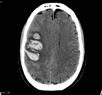
Venous Sinus Thrombosis Image

Venous Sinus Thrombosis Image

Image source: radiopaedia.org

The presence of blood clot in the venous sinuses prevents venous blood from draining from the brain. These may lead to congestion in the veins and may lead to rupture once the pressure in very high. Rupture leads to hemorrhage and subsequently, stroke. Stroke causes severe hypoxia of the brain cells leading to permanent damage. This condition is usually life-threatening.

Venous sinus thrombosis can occur at any age and even among newborns. Its occurrence in newborns is usually apparent during the first two months of life.

Types of Venous Sinus Thrombosis

Cerebral venous sinus thrombosis
This involves the presence of thrombosis in the deep cerebral veins. It is a rare occurrence, which constitutes 30% of all cases of venous sinus thrombosis.

  • Dural Venous Sinus Thrombosis – This involves the formation of clot on the superficial veins in the dura mater. It is also a rare occurrence along with the cerebral veins.
  • Cortical Venous Sinus Thrombosis – The thrombosis is located on the cortical veins, where capillaries in the brain merge.
  • Saggital Venous Sinus Thrombosis – The saggital venous sinuses are a common site for venous sinus thrombosis. Superior saggital sinus thrombosis is most likely associated with Cerebrovascular Accidents or Stroke.
  • Transverse Venous sinus thrombosis – Transverse venous sinus thrombosis is more uncommon than saggital thrombosis, however, the occurrence of which most likely cause increase in the intracranial pressure.

Venous Sinus Thrombosis Symptoms

Symptoms of venous sinus thrombosis are usually a sign of increased intracranial pressure. These include:

  • Headache -Headache is present in up to 90% of cases. It can be in the form of thunderclap headache or the presence of a sudden headache in just a period of minutes. It is also characterized as new onset headache that is persistent. Headaches may also in the form of cluster headache, which involves pain in more than one area of the head. The headache is usually worsened during coughing, lying, or straining. There may also be presence of severe headache in the morning.
  • Vomiting – The increased intracranial pressure also causes projectile vomiting because of the increased pressure that compresses the vomiting center, which is the medulla oblongata.
  • Pulsatile tinnitus – Pulsatile tinnitus is present because of the pressure in the otic nerve.

Tinnitus Image

Pulsatile tinnitus as  a symptom of Venous Sinus Thrombosis

  • Neck stiffness – Neck stiffness may also be present because of too much pressure in the venous channels in the brain, which radiates to the neck area.
  • Papilledema – Papilledema is the swelling of the optic disc as a result of increased intracranial pressure. It causes visual obscurations or blind spots.

The symptoms of venous sinus thrombosis do not appear in some patients. The most apparent symptom is new onset headache.

Causes and Mechanism

The causes for children and infants are usually different from the risk factors of adults. These include:

Causes in Infants and Children:

  • Sickle cell anemia – The presence of this condition causes early hemolysis of red blood cells. When these are hemolyzed in the venous sinuses, it can cause blood clotting in the area.
  • Problems with blood clotting – Newborns may have congenital defects of the blood clotting mechanism causing severe blood clotting that can obstruct the venous sinuses.
  • Dehydration – The presence of dehydration causes the blood to become more viscous, thereby increasing risk for clotting.
  • Head Injury – Head injury during childhood may cause bleeding on the venous sinuses and lead to blood clots in the area.
  • Infections in mothers during pregnancy – Mothers may experience certain infections during pregnancy and may lead to spread to the developing fetus. The infection may travel to the brain of the fetus causing blood vessel injury and subsequently blood clots.

Causes in Adults

  • Pregnancy and puerperium – Pregnancy has been one of the most common causes of VST in adults. Pregnancy related conditions include increase maternal age, cesarean delivery, hyperemesis and maternal infections
  • Use of oral contraceptive pills – Contraceptive pills increases the risk for abnormal blood clotting that may happen on the venous sinuses.
  • Cancer – Malignancies in the brain or metastatic conditions can also cause blood clots in the venous sinuses as a result of damage to the intima of the blood vessels caused by tumors.
  • Collagen vascular diseases – Diseases such as systemic lupus erythematosus may also lead to VST because of risk of blood clots in the brain.
  • Inflammatory bowel disease – Chronic inflammatory conditions such as Crohn’s disease increase the C-reactive protein in the blood, which stimulates blood clotting because of trauma.
  • Obesity – Obesity may also cause VST because of elevated cholesterol levels in the body.
  • Intracranial hypotension – The presence of hypotension in the area leads to decreased venous blood flow. A sluggish blood flow can result in clot formation.

Diagnosis

The diagnosis of Venous Sinus Thrombosis uses the following tests:

Venography

Venography is done by injecting a contrast medium in the veins in the cranium with series of imaging tests to determine any obstruction of blood flow. Venography is usually assisted by the use of CT scan (CT venography) or MRI (MR venography). This test is usually done when absence of arterial obstruction is seen.

Lumbar puncture

Lumbar puncture is made to assess the opening pressure in the CSF that is increased when venous sinus thrombosis is present.

Treatment

Treatments of Venous Sinus Thrombosis include:

Anticoagulation

Anticoagulation therapy with the use of heparin and warfarin is given to prevent the formation of additional clots. These medications are not given to infants and children because of risk of severe hemorrhage.

Thrombolytic therapy

Thrombolytic drugs are those that directly dissolve a blood clot. This is usually given to eradicate the thrombus, which causes ICP elevation. Example of this drug is streptokinase.

Lumbar Puncture

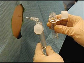
Lumbar puncture is also employed to reduce the intracranial pressure. This can be done along with the diagnostic lumbar puncture by aspirating the CSF.

Lumbar Puncture Image

Lumbar Puncture Image

Anticonvulsants

Anticonvulsants are given to prevent seizures as a complication of VST.

Diuretics

Just like in any other increased ICP conditions, diuretics are given to reduce the CSF in the CNS.

No responses yet

Condylomata Acuminata – Pictures, Cure, Symptoms, Treatment, Causes

Apr 17 2012 Published by under Diseases & Conditions

What is Condylomata Acuminata?

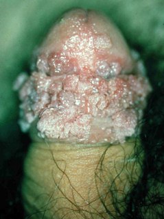
Condylomata Acuminata or so called Genital Warts is a type of sexually transmitted disease that is caused by a human papillomavirus. This disease is spread through sexual contact, its either oral sex, genital or anal sex. Genital Warts are flesh colored, cauliflower in shaped that can be seen in both male and female.

This disease is considered as one of the most common sexually transmitted disease. Age group that is usually had this disease is between 17-33 years of age.

Condylomata Acuminata Image

Condylomata Acuminata Image

Symptoms of Condylomata Acuminata

The warts look like flesh colored spots, looks like a cauliflower and it can be elevated or flat and it is usually painless

In females:

  • It can be located at the inside of the anus or vagina
  • It can be also located in outside of vagina or anus or it can be present in the nearby skin
  • It can go up to the cervix inside the body

In males:

It can be located at:

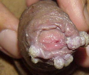
  • PenisGenital warts Image

Genital warts on penis

Image source: img.medscape.com

  • Scrotum
  • Groin area
  • In thigh area
  • It can also be located inside of the anus or
  • Around the anus area
  • In can also occur in other places like:
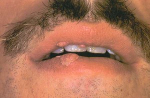
  • Lips
  • Mouth
  • Throat and
  • Mouth
  • Other symptoms that is related in Genital Warts are:
  • There is an increased in dampness in the genital area
  • There will also be an increase in vaginal discharge
  • There will be also a sign of vaginal itching
  • Sometimes bleeding can occur during or after sexual intercourse
  • Pain especially if the wart is located at cervix and vagina
  • There will be a visible tiny papules in the males penis
  • There is a possible burning sensation in the genital and anal region especially if the case is already severe.

Causes of Condylomata Acuminata

  • This disease is main caused by Human Papillomavirus (HPV)
  • Important facts about Human Papillomavirus
  • It is spread through sexual contact
  • It will take a year for you to notice the presence of genital wart
  • Not everyone who has a direct contact with the disease can have or can be acquired by them
  • You can easily get Genital warts and be able to spread them quickly if:
  • You have multiple sexual partners
  • You don’t have any idea that your partner is affected by a sexually transmitted disease
  • You are already sexually active at an early age
  • You smoke
  • You take alcohol
  • You have a present viral infection that weaken your immune system

Diagnosis

  • Patient history (Medical and Sexual History)
  • Physical Examination to spot the signs and symptoms of having genital warts. It will include the observation of the genital area and pelvic region as well as presence of warts in mouth and tongue
  • The medical professional will also do Colposcopy. It is where the doctor will used a lighted magnifying device that will be inserted in order to have a better visualization of the vagina to help the medical professional to check any signs and symptoms of having genital warts.
  • The medical professional advised a woman to take a Pap smear examination. Pap smear will help medical practitioner to check for any abnormalities. The medical professional will scrape some cells that are present on the uterine cervix.
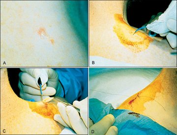
  • Biopsy is also recommended where the medical professional will scrape some of the part of the wart in order to determine the exact cause of the disease.

Biopsy Image

Biopsy to diagnose Condylomata Acuminata

Treatment of Condylomata Acuminata

Medicines

  • Imiquimod (Aldara)
  • Podophyllin and podofilox (Condylox)
  • Trichloroacetic acid (TCA)

Surgery

  1. Surgical Excision in this type the medical professional will cut out the wart and the patient is in local anesthesia
  2. Laser Therapy is a therapy where an intensive beam of light is used to destroy the present wart
  3. Electrocauterization it is where the medical professionals will use electric current to destroy the wart and usually the patient is on local anesthesia.

Prevention

  • Ask your Doctor about the vaccine against Human Papillomavirus
  • Use of protective devices such as condom
  • Avoid direct contact with the virus that causes Condylomata Acuminita

Prognosis

The prognosis of disease depends on the attitude of the person who has it. Usually sexually active woman is the one who is affected by Human Papillomavirus and usually it goes away on its own. While most men never develop any symptoms like infection, they can still be able to pass it to their sexual partner.

Condylomata Acuminata Pictures

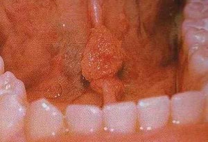
 Condylomata Acuminata lower lip

Condylomata Acuminata of lingual frenulum

Condylomata Acuminata

Source – siamhealth.net

Condylomata Acuminata anal

No responses yet

« Prev - Next »

© 2017 eHealthWall.com. All Rights Reserved. Privacy Policy
This website is for informational purposes only and Is not a substitute for medical advice, diagnosis or treatment.