Archive for May, 2012

Hyperemesis Gravidarum – Definition, Treatment, Causes, Diet, Symptoms

May 14 2012 Published by under Pregnancy

What is Hyperemesis Gravidarum?

Hyperemesis Gravidarum is a kind of disease that occurs during pregnancy where the mother experience excessive and persistent nausea and vomiting. Having this type of condition, it can lead the mother’s to be dehydrated.


hyperemesis

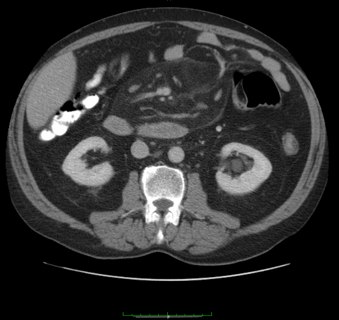
Hyperemesis Gravidarum Image

Hyperemesis Gravidarum Symptoms or signs of start of morning sickness:

  • The pregnant woman will experience excessive nausea normally during the first trimester of pregnancy.
  • They also experience excessive vomiting that can lead to dehydration.
  • Weight loss that is due to excessive vomiting that makes the pregnant mother also not to keep up with eating of food.
  • The pregnant mother with this disease will experience fatigue.
  • They also feel dizziness.
  • Dehydration that will lead to feeling of thirst, headache, confusion and reduced urination.
  • Low blood pressure and increased heart rate is also one of the symptoms that the pregnant woman with this type of disease will experience.

Hyperemesis Gravidarum Causes

The causes of Hyperemesis Gravidarum are: (1) increased in human chorionic gonadotropin (2) increase in estrogen level (3) multiple pregnancy (4) hydatidiform mole or also called as h-mole.

Risk Factors that contribute in Hyperemesis Gravidarum

  • The mother already experience previous Hyperemesis Gravidarum
  • The mother is overweight
  • Having a multiple pregnancy or formation of the Hydatidiform mole.
  • First pregnancy of the mother
  • The mother is being pregnant in young age
  • The mother has no completed pregnancies
  • There is a family history like a mother or a sister that experience the same thing
  • Unplanned pregancy

Diagnosis

Patient History

That will contribute in diagnosing Hyperemesis Gravidarum to pregnant woman.

Assessing Patient's History Image

Assessing Patient’s History to determine the cause of Hyperemesis Gravidarum

Physical exam

To see visible signs that can be related in diagnosing Hyperemesis Gravidarum.

Laboratory test

  • Like checking Hematocrit and Ketones Level in the urine to check signs of dehydration.
  • Liver Enzyme and Bilurubin that if there shows an increase in Transaminase level there is a possibility that it occur to the mother’s with Hyperemesis Gravidarum and to eliminate other possible disease that affects the liver and bilurubin enzyme.
  • Checking for Thyroid Stimulating Hormone that can trigger the vomiting and nausea of the pregnant mother.
  • Urine culture to check signs of infections that is common to occur in pregnant woman.
  • Ultrasound that will help to detect if you have twins or if you have any present disease in the uterus like H-mole (hydatidiform mole) that causes your excessive nausea and vomiting.

Hyperemesis Gravidarum Treatment

Intravenous Therapy

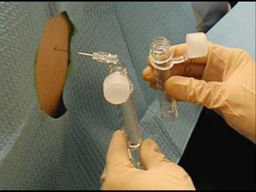
  • For pregnant woman, hydration is needed. In the case of experiencing Hyperemesis Gravidarum, where there is an excessive vomiting that can lead to dehydration, giving intravenous fluids is a must to prevent problems such as dehydration.
  • It will help to supply the electrolytes that are lost due to recurrently vomiting that will lead in deficiency

Medications

  • Anti-emetic drugs that is used to prevent or avoid nausea and vomiting.
  • Vitamin B6 that is known for its effectiveness to decrease the feeling of nausea during pregnancy.
  • Medication: Benedictin or Dicletin : combination of Vitamin B6 and doxylamine : best medication to treat hyperemesis gravidarum : anti-nausea medication
  • Medication: Zofran or Ondansetron : helps prevent nausea and vomitting

Total parental Nutrition

  • If the pregnant woman experience a severe form of Hyperemesis Gravidarum, the medical professionals will advise the mother to have this kind of treatment where the food and the nutrients needed in pregnancy is given through intravenous line.
  • Nasogastric Tube Feeding – a kind of tube that is inserted from the nose to the stomach : it is the commonly used procedure to give the needed nutrition by the mother.

Nasogastric tube in pregnant woman with Hyperemesis Gravidarum

Nasogastric tube in pregnant woman with Hyperemesis Gravidarum

  • Percutaneous Endoscopic Gastrostomy – is a surgical type procedure that the tube is inserted through from the abdomen to the stomach.

Other measures

  • Eating and drinking foods that are clear and bland type of foods that are rich in carbohydrates.
  • Eating in small frequent meals to avoid too much weight loss and dehydration
  • Ginger is known for its useful properties in treating nausea in pregnant woman but not all women that experience hyperemesis gravidarum that the ginger is effective.
  • Medical professionals advised pregnant woman to have a complete bed rest during the treatment period that might offer comfort.

Prognosis

Pregnant woman who has signs of excessive vomiting and nausea should go immediately to medical professionals for check-up and early indentifying the cause of the symptoms present to avoid further problems that will risk the health of the mother and the baby. This condition not often causes serious problems between the mother and the baby.


No responses yet

Meningococcemia – Rash, Symptoms, Treatment, Pictures, Causes

May 11 2012 Published by under Diseases & Conditions

What is Meningococcemia and what causes bacterial meningitis?

Meningococcemia is a kind of infection that affects the person’s bloodstream which may be due to the bacteria called as Neisseria meningitidis, which is a gram negative kind of bacteria. The bacteria is contagious in nature and has the ability to spread from one person to many through respiratory mode via the secretions or droplets.

Although meningococemia may be etioligally due to variously wide class of disease. The severest form of this syndrome is the neiserial meningitis which is often the most life-threatening disease.This kind of disease, as mentioned earlier, which is caused by neisseria meningitidis is that when one has this condition there is a great chance that it will progress into meningitis and it may also result inot dermatologic problems.

Meningococcemia Image

A child who has Meningococcemia

Image source: wikimedia.org

Meningococcemia Pathology

When a person is positive for menigococcemia, he or she will have the bacteria, neisseria meningitidis, which attaches to the respiratory mucosa and are taken by the phagocytes, which functions as the protector of the endothelial cells and capsule. When the bacteria reaches to this point, the bacteria will then release endotoxin which will then damage the vessels which will result to the symptoms that will be discussed further as we go along.

Meningococcemia Symptoms

The patient with meningococcemia will manifest the following symptoms:

  • Arthralgia
  • Myalgia
  • Fever
  • Petechia
  • Bullae
  • Hemorrhagic lesions
  • Maculopapular rash lesions
  • Stellate pupura with having a gray hue* late sign
  • Anxiety
  • Irritability
  • Nausea
  • Fatigue
  • Chills
  • Sore throat
  • Vasculitis
  • Muscle pain
  • Headache
  • Shock * late sign
  • The level of consciousness changes * late sign
  • Myocarditis* late sign

These are the most common symptom associated with this kind of disease condition.

Meningococcemia Pictures

Stellate pupura with having a gray hue (rash)

Image source: medlibes.com

Meningococcemia rash

Meningococcemia Causes

As mentioned earlier, the cause of this delibitating disease is due to neisseria meningitidis or otherwise known as meningococcus. This disease condition may also be called as meningococcal bacteremia. What happens is that the bacteria before it attacks to the entire body, will lodge in the person’s uppermost portion of the respiratory tract silently or asymptomatically. The bacteria is contagious and will affect and spread to one person to another person via respiratory mode in the form of secretions such as droplets.

Although, some persons will be affected and will show signs associated with the disease condition. However, some who are affected with the disease condition may not show signs of the disease and are often tagged as carrier of the disease condition. This carrier persons may have the ability of spreading the disease to others.

Meningococcemia Diagnosis

The physician, who are the ones capable of diagnosing the patient with meningococcemia, would suggest the patient to undergo examinations such as:

  • Physical examination
  • Medical history examination
  • Blood culture examination
  • Clotting examination
  • Complete blood count examination that has differential
  • Urinalysis
  • Skin biopsy
  • Lumbar puncture examination

The kind of examination done with the patient who are suspected to have meningococcemia will vary from person to person. Yet, the ones mentioned above are the possible examination that the patient must undergo to confirm the disease condition.

Meningococcemia Terminology

Fulminant Meningococcemia

The fulminant meningococcemia is a kind of disease condition that is rarest under the classification of menigococcal disease condition which will result to often a fatal form of the disease condition. The deterioration of this kind of disease classification may also lead to the failure of both respiratory and heart which may result to hypotension.

Chronic Meningococcemia

When a person has a chronic form of meningococcemia, it means to say that the symptoms are worsening and recurring. When one reaches this point, there will be a risk for sepsis if the condition is not well tolerated and especially if the body does not anymore respond to the treatment being given. It is a rare kind of classification of meningococcemia which occurs more often in adults. This kind of disease, because of the fact that is resembles to other forms of diseases such as vasculitis and the like thus there is a need that there should be a strong suspicion of the disease to be able to come up with the right treatment for the disease condition.

Meningococcemia Treatment

When one is diagnosed with this kind of disease condition, which is known to be a medical emergency, there should be a surveillance of the progression of the disease condition. The patients are often admitted to the intesive care unit to be able to have a close monitoring of the patient having the disease condition. It is only in the intensive care unit, wherein there is one is to one ratio of the patient to his or her nurse, which is an ideal set up especially with this disease condition. In addition to that, since this disease can be spread via respiratory secretion thus the patient has to be in isolation, specifically in respiratory isolation to prevent the spread of the infection to others. Other treatment may be done, such as:

  • IV fluids
  • Wound care
  • Replacement of platelets or clotting factors
  • Antibiotics
  • Breathing support such as mechanical ventialtion or oxygen therapy
  • Drugs for low blood pressure
  • Corticosteroid drugs
  • Surgeries can be done like:
  • Periocardiocentesis for patients with pericarditis with a complication of tamponade
  • Amputation for arterial occlusion

When the patient is treated early, there is a high chance that the patient will have a good prognosis and he will survive this disease condition. Hence, it is advisable that that the patient should consult a doctor as soon as possible whenever symptoms will arise. However, when the person reaches the late stages and shock may develop the chances for survival or living is poor and the outcome of the treatment will be a little less certain.

On the otherhand, with persons who are at risk for the disease condition, there is a need to have to undergo antibiotic therapy and vaccination, which is specially done in children and in older persons to prevent the spread of the disease condition. You need to talk with the doctor with the options presented in your area.

No responses yet

Mesenteric Panniculitis – Definition, Symptoms, Causes, Treatment

May 06 2012 Published by under Diseases & Conditions

What is Mesenteric Panniculitis?

Panniculitis refer to the inflammation of the fatty deposits or adipose tissue that can be seen most particularly in the abdominal area. Mesenteric Panniculitis is a rare inflammatory disease that affects the subcutaneous adipose tissue of the mesentery or the small bowel area that is characterized by blockage to the small intestine. It was first referred to as retractile mesentery by Jura in 1024 and was renamed as mesenteric panniculitis by Odgen in 1960.

There have been so many terms being used to refer to the disease, but today it was classified into one disease of two categories; the mesenteric panniculitis and retractile mesentiritis. It is known as mesenteric panniculitis if the inflammation and fat necrosis is more than the fibrosis, whereas retractile mesentiritis if fibrosis dominates.

Also, another distinguishing characteristic of mesenteric panniculitis if the presence of mesenteric lipodystrophy by fat necrosis, chronic inflammation and retractile mesenteritis by fibrosis.

There is a higher of incidence report in middle aged males compared to females, and it is common in Caucasian males. Studies also show that as person ages, the risk to have possibly had the disease also increases. On the contrary, there is no incidence yet reported on the occurrence of the disease to pediatric patients, maybe because they have less fats, which is the major contributing factor of the disease, as compared to adults.

Mesenteric Panniculitis Image

Ct Scan Imaging to depict presence of Mesenteric Panniculitis

Symptoms and Signs

Because it affects most on the abdominal area, the symptoms are also related to the affected system and may last for some time or even more than a year. Although it appears to be showing no symptoms at all, its manifestation differs between individuals and cases. The disease may present symptoms such as:

  • Abdominal pain of varying intensity
  • Loss of appetite
  • Back pain
  • Nausea
  • Fever
  • Weight loss
  • Abdominal fullness
  • Palpable abdominal mass
  • Constipation or diarrhea
  • Passage of blood in the rectum

In some instances, the patient may not show any physical manifestation unless with the aid of some diagnostic assessment. Other patient also report to experience to have jaundice, acute abdomen and abdominal obstructions. With the different manifestation of the disease will also entail several illnesses that have to be taken into consideration.

Mesenteric Panniculitis Causes

There is no known specific cause of the disease and until today it is still unclear for those who are in the medical profession. However, there are some predisposing factors that can lead to the development of the disease.

Mesenteric panniculitis is believed to affect to those people who undergone surgery. Tumor and other inflammatory condition affecting the abdomen cannot be discounted in contributing to the occurrence of the disease. Autoimmune diseases like SLE, Thyroid disease and other related condition are also said to cause the disease.

The condition is also said to be linked to the progression of the disease are mesenteric arteriopathy, chemical, thermal and drug injuries, avitaminosis, pancreatitis, leakage of bile or urine, and bacterial infection.

There are also some other factors like gallstones, heart diseases, peptic ulcer, cirrhosis, abdominal aortic aneurism, are said to also contribute to cause the disease. There are also some studies that claim of the significant relationship of tobacco smoking to mild mesenteric panniculitis.

On the other hand, Retractile Mesentiric is associated with those diseases that have something to do with fibrose tissue. These disease are related to malignant disease such as lymphoma, melanoma. Hodgkin’s disease, renal cancer cells, and other types of cancer.

Mesenteric Panniculitis Diagnosis

Because of the extensive effect of the disease, the assessment and diagnosis also differ depending on the presenting factor it has of the individual.

Abdominal Computed tomography is believe to be the most preferred in finding the abdominal mass present on the body. This will also somehow help in identifying the presence of the “fat ring” sign that shows the presence of fat deposits and the “tumor pseudocapsule”. But, CT scan does not help in the diagnosis of the disease since they are thought to have no clear impressions. The presence of the disease in the CT scan appears to be soft tissue mass being covered by a mesenteric vessel that later progress into collateral vessels.

The presence of fat around the mesenteric vessels was referred to as the fats ring sign. This feature makes it differs to other condition of the same origin such as lymphoma, carcinomatosis or carcinoid tumor. Another thing that also needs to be considered it the appearance of the mesentery in the CT scan, it’s misty appearance are also the same characteristic that are being displayed by haemorrhage, edema or lymphoma.

A more definite diagnostic tool is the MRI. It reveals the existence of fibrous capsule that cannot be identified by other diagnostic tools. Still surgical biopsy remains to be the almost precise and acceptable diagnostic evaluation to determine the presence of Mesenteric Panniculitis since they can provide a complete histologic analysis.

Histological examinations are also significant in confirming the occurrence of the disease but they are not as definitive as the radiologic examinations. Blood works will reveal a normal value.

Histological Examination Image

Histological Examination to diagnose presence of Mesenteric Panniculitis

There are some radiologic examinations that can be used to identify the presence of Mesenteric Panniculitis, yet their reliability is still in question.

Mesenteric Panniculitis Treatment

The treatment of Mesenteric Panniculitis depends on the severity of the condition and there is no definite or universally accepted form of treatment for the disease. Correct diagnosis must be undertaken before doing any treatment since looking for the exact finding of the condition is often difficult.

The most common therapy is with the use of steroids, colchicine, tamoxifen, cyclophosphamide, progesterone, antibiotics and a lot more. Prednisone is known to have a favourable effect and delivers the quickest response among corticosteroids. The effects of these medicines have different rate of success and its effectiveness varies also among individuals.

If medication therapy be unsuccessful, surgical treatment might be done if it entails a life-threatening situation occurs and a colostomy may be created, although there are some medical practitioner who do not prefer for such option.

Their reason of opposing to surgery is that it might aggravate the condition. In benign cases, surgical resection has proven to cause progression of the condition thus leading to death of the patient. If surgical treatment is to be pursued, then a medical history review is encourage to prevent other complications in occurring.

Surgical Resection to diagnose presece of mesenteric panniculitis image

Surgical Resection to diagnose presence of Mesenteric Panniculitis

On the other hand, radiation therapy are said to be not useful and no proven effect in the resolution of the disease.

Radiation Therapy Image

Radiation Therapy to treat Mesenteric Panniculitis

Mesenteric Panniculitis Prognosis

The overall prognosis of the disease is good, although there are some very few instances of recurrence. On the other hand, it should also be noted that the success of the treatment is inconsistent and depend on the severity of the condition.

There are some incidents that because the patients are asymptomatic, they will seek treatment if conditions are already classified as malignant.

No responses yet

Optic Nerve Hypoplasia – Definition, Treatment, Causes, Symptoms

May 03 2012 Published by under Diseases & Conditions

What is Optic Nerve Hypoplasia?

Optic nerve hypoplasia is a condition involving the underdevelopment of the optic nerves that commonly occurs as a result of congenital optic nerve anomaly. In optic nerve hypoplasia, the optic nerve axons have not developed properly leading to an abnormally small optic disc. The optic nerve is responsible for transmitting the signals of vision from the retina to the brain. The optic nerve usually consists of up to 1.2 million of optic nerve fibers. In optic nerve hypoplasia, there are fewer nerve fibers leading to problems in the vision.

Optic Nerve Hypoplasia

Optic Nerve Hypoplasia Image

Image source: njms2.umdnj.edu

Optic nerve hypoplasia is more commonly related to brain malformations, developmental delay as well as endocrine problems or hormone deficiency. Aside from the problems in the optic nerve, there are also problems relating to anomalies in the hypothalamus, maldevelopment of the pituitary gland, absence of septum pellucidum and agenesis of the corpus callosum. Due to these, patients with optic nerve hypoplasia are also prone to hormonal deficiencies and developmental delays.

Optic nerve hypoplasia has become the leading cause of infant blindness in Sweden, overtaking the position from retinopathy of prematurity. Optic nerve hypoplasia may affect all races, but is lesser prevalent in Asian populations.

Optic Nerve Hypoplasia Symptoms

Optic nerve hypoplasia causes symptoms in the vision as well as symptoms related to the accompanying problems in the brain. These include:

Vision problems

Children with ONH suffer from strabismus or the inability to align the eye simultaneously or nystagmus or the involuntary eye movements. Vision problems may be bilateral or unilateral. Children with unilateral vision problem have better vision. Vision problems tend to improve during the first few years of the child. Vision problem also do not result in decline or progression.

Strabismus Image

Strabismus as one of the symptoms of Optic Nerve Hypoplasia

Image source: babble.com

Hypothalamic symptoms

Because of problems in the hypothalamus, children often lead to problems in the regulation of the pituitary gland. The pituitary gland is responsible for the regulation of the hormones in the body. Problems in the hypothalamus may lead to poor functioning of the pituitary gland or hypopituitarism. Hypopituitarism is often present in up to 80% of patients with ONH. As a result, there is poor function leading to lack of growth hormone, adrenal insufficiency, hypothyroidism and diabetes insipidus. Symptoms depend on the hormone affected. These include:

  • Growth Hormone Deficiency
  • Short stature
  • Hypoglycemia
  • Seizures
  • Jaundice
  • Delayed dentition
  • Micropenis in boys
  • Hypothyroidism
  • Hypothyroidism may lead to mental retardation when left untreated because of poor metabolism and development of the brain. Hypothyroidism manifests as:
  • Cold intolerance
  • Water retention
  • Rapid weight gaining
  • Adrenal Insufficiency
  • Precocious puberty because of problems in sex hormones
  • Obesity
  • Inability to cope with stress because cortisol deficiency
  • Diabetes insipidus
  • Excessive urination
  • Dehydration
  • High sodium levels in the body

Other problems involving hypothalamic affectation involve problems in sleep, feeding and regulation of the body temperature. Because of these, children often exhibit overeating and obesity or reduced food intake that may or may not lead to weight loss. Children also suffer from abnormal sleep wake cycle leading to behavioral problems.

Neurologic problems

The presence of malformations in the brain causes developmental delay such as motor delays, which affects up to 75% of cases and communication delay, which affects up to 44% of children.

Optic Nerve Hypoplasia Causes

Optic nerve hypoplasia is related to several prenatal and perinatal risk factors. These include:

  • Young maternal age – Mothers who are young have been related to the development of optic nerve hypoplasia.
  • Primiparity – Infants who are first born usually suffer from the condition.
  • Increased number of cesarean delivery among mothers
  • Gestational vaginal bleeding
  • Preterm labor
  • Low maternal weight gain
  • Weight loss during pregnancy.

All these factors are associated with the development of the fetus leading to optic nerve hypoplasia.

Optic Nerve Hypoplasia Diagnosis

The diagnosis of ONH involves opthalmoscopic examination that indicates small optic nerve. The DM:DD ratio is also used to ascertain diagnosis. DM refers to Disk to Macula, which is the distance of the center of the disc to the macula. DD refers to the Disc Diameter. When the ratio is greater than 3, there is suspicion of ONH. However, when the ratio is greater than 4, ONH is definite.

Optic Nerve Hypoplasia Treatment

Optic nerve hypoplasia has no treatment because of permanent damage of the optic nerve. However,optic nerve hypoplasia treatments are available for the symptomatic relief of patients. These include:

  • Hormone Replacement Therapy – The presence of hypopituitarism may require hormone replacement to manage symptoms of hypopituitarism as wells as regain the normal functioning of the body. Hormone replacement may include growth hormone therapy, thyroid hormone replacement, estrogen and testosterone therapy and others. These treatments reverse the effects of hypopituitarism in order for the client to live normally.
  • Physical and occupational therapy – These therapies are also needed to help patients achieve optimum functioning and address motor delays. These therapies are started as soon as possible to allow children to minimize functional disabilities.
  • Speech therapy – Speech therapy is also beneficial to assist the child in communication as a result of communication delays. Speech therapy may start as early as infancy when the child learns how to talk.
  • Melatonin administration – Melatonin is also given to enhance the sleep pattern of children. Melatonin given at night adjusts the circadian rhythm of children that have been affected by hypothalamic dysfunction.
  • Eye patching – Patching of the good eye is done to improve the vision of the affected eye in the presence of strabismus. This technique allows the better use of the affected eye. Eye patching is only done in cases where the vision is known to improve in both eyes.
  • Optic Nerve Hypoplasia Surgery – Surgery for children involves the alignment of the eyes in children with strabismus. The surgery involves the repair of the muscles that control the eye movement. Eye surgery is done when the child already has developed visual acuity, which is commonly achieved after three years of age. However, the eye surgery only involves correction of the eye deformity and it does not improve the vision.

After treatment, the prognosis of ONH is variable. Patients may have near normal vision and some may have legal blindness. Patients may also suffer from intellectual disabilities, despite treatment.

No responses yet

Pseudotumor Cerebri – Symptoms, Causes, Treatment and Diagnosis

May 01 2012 Published by under Diseases & Conditions

What is Pseudotumor Cerebri?

Pseudotumor cerebri or PTC is a disorder in the neurologic system involving an increase in the intracranial pressure without any cause such as diseases and tumor.

Intracranial pressure (ICP) is the pressure maintained inside the cranium around the brain. Pseudotumor cerebri is also called benign intracranial hypertension (BIH) or Idiopathic intracranial Hypertension (IIH).

PTC can occur in all age groups and is common in women. The incidence of PTC is about one in every 100,000 population. The disorder occurs in women 20 to 45 years of age. Women have a very high risk of developing the condition and are up to eight times more prone for PTC than men.

Specifically, women who are obese or overweight are predisposed to pseudotumor cerebri. In children, the occurrence of PTC is equal in males and females.

Symptoms of Pseudotumor Cerebri

The symptoms of pseudomotor cerebri are rooted with increased intracranial pressure. These include:

  • Headache – Headache is the most common symptom of the condition and it appears to more than 90% of cases. Headache is characterized as throbbing, generalized and worsens in the morning. It can also be aggravated by Valsalva maneuver, coughing, straining on stools and sneezing. The pain may radiate to the shoulders and neck.
  • Nausea and Vomiting (projectile) – The increased pressure compresses the brain because the cranium is a closed system. When the brain is compressed, the medulla oblongata (vomiting center) is affected.
  • Pulsatile Tinnitus – Tinnitus is the presence of ringing of the ears and is seen in up to 87% of patients. The tinnitus is synchronous with the pulsetinnitus Image

Tinnitus as one of the symptoms of Pseudotumor Cerebri

Image source: tinnitusdx.com

  • Loss of senses – The senses may also be affected specifically the sense of smell because of possible swelling, traction and compression of the cranial nerves.
  • Lack of coordination and balance – The increased ICP also compresses the cerebellum, which is responsible for balance and coordination.
  • Double vision – People with abducens nerve palsy, damage to the sixth cranial nerve, may result to double vision because of the compression of the cranial nerves that pulls the eye outward.
  • Facial muscle weakness – The facial nerve may also be affected causing problems on facial expression and movements.
  • Papilledema – The increased ICP spreads the pressure on the optic nerve located on the eyes causing the optic disc to swell. Papilledema causes blind spots or visual obscurations. Papilledema is diagnosed using opthalmoscopy.

Pseudotumor Cerebri

Papilledema due to increased intracranial pressure

Image source: webeye.ophth.uiowa.edu

Causes of Pseudotumor Cerebri

The cause of pseudomotor cerebri is unknown, which made it as idiopathic. Pseudotumor cerebri is diagnosed when other conditions or causative factors are not present to increase the intracranial pressure. Increased intracranial pressure, which is caused by certain factors are termed as secondary intracranial hypertension.

The cause of pesudotumor cerebri may be linked with the Monro-Kellie rule that any increase in the brain tissue, CSF and blood may lead to increased intracranial pressure. However, the exact root cause is not known.

Obesity and being overweight were linked to the occurrence of the condition because of increase in venous pressure.

Diagnosis of Pseudotumor Cerebri

Diagnosis of pseudotumor cerebri is made when other conditions are not present to increase ICP. The specific diagnostic tests used are:

Brain Scan

A brain scan with the use of MRI or CT scan is used to rule out other causes of the disorder. MRI findings and CT scan results may show normal scans because of the absence of any tumor and obstruction. However, patients may exhibit pituitary gland flattening or empty sella sign as a result of increased pressure inside the cranium.

Brain Scan Image

Brain scan images to show flattening of pituitary gland

MR Venogram

This utilizes magnetic resonance imaging while contrast medium is injected on the veins in the brain. This test is used to determine any occurrence of cerebral venous sinus thrombosis or venous obstructions. This test is done for patients with atypical conditions to further evaluate the occurrence of any probable cause.

Lumbar Puncture

Lumbar puncture is done to measure the pressure in the CNS. The CSF may also be examined for presence of tumor or infection in the CNS, which may result to increased ICP. Proponents suggest that lumbar puncture should be taken while the patient is on a side lying position because sitting position may cause false positive increase in opening pressure.

The criteria for the diagnosis of poseudotumor cerebri use the Dandy criteria for the disease. These include:

  • Presence of symptoms of elevated ICP
  • No localizing signs, but may present abducens nerve palsy
  • Normal brain scan findings
  • Patient is alert and awake
  • Opening pressure of greater than 25 cmH20 upon lumbar puncture with absence of abnormal components or cytological composition of the cerebrospinal fluid
  • No other possible explanation for the pressure increase

Lumbar tap

Lumbar puncture to measure increasing pressure in the brain and examine possible CNS tumor or infection.

Treatment of Pseudotumor Cerebri

The treatment of pseuditumor cerebri is focused on decreasing the intracranial pressure because no known cause can be treated. Treatments also prevent the development of visual loss as a result of papilledema. Pseudotumor cerebri can be resolved after treatment, but some may progress chronically. Some patients also go into remissions with the possibility of a relapse. The treatments involved include:

Lumbar Puncture

Aside from using it as a diagnostic test, lumbar puncture is done to remove or drain CSF in order to decrease the intracranial pressure. The procedure involves the insertion of a thin needle through the spine and the CSF is aspirated afterwards. Lumbar puncture may be able to relieve increased ICP immediately and patients may not require additional treatments. Lumbar puncture can be repeated according to the need, but this requires caution because microorganism can enter the spinal fluid and cause infections. Lumbar puncture is also an emergency measure for progressive vision loss as a result of papilledema.

Diuretics

Diuretic such as acetazolamide (Diamox) is given to patients with increased ICP. This medication inhibits carbonic anhydraze, thereby reducing the production of CSF. Diamox can cause potassium loss through the urine so monitoring of potassium levels is essential. Furosemide may also be given when patients do not tolerate the former drug.

Analgesics and Steroids

Analgesics may be given to relieve headache. Corticosteroids are also given to reduce cerebral edema and reduce ICP.

Surgery

Surgery may be employed when increased ICP is not treated with conventional therapies. This includes procedures such as optic nerve sheath decompression, fenestration and ventriculoperitoneal shunting. Optic nerve sheath decompression and fenestration decreases the tension in the optic nerve to prevent vision loss. VP shunting is needed to allow more efficient elimination of excess CSF in the ventricles in the brain.

Prognosis

Pseudotumor cerebri does not affect the life expectancy. Patients can be treated completely as long as the ICP is decreased. The condition can eventually lead to vision problems when not treated promptly.

Complications

The main complication of the condition is vision loss as a result of prolonged papilledema. Vision loss may affect up to 25% of patients.

ICD-9 Code

The ICD-9 code of pseudotumor cerebri is 348.2

No responses yet

« Prev

© 2017 eHealthWall.com. All Rights Reserved. Privacy Policy
This website is for informational purposes only and Is not a substitute for medical advice, diagnosis or treatment.