Archive for April, 2016

Perioral Dermatitis – Children, Treatment, Symptoms, Causes, and Diet

Apr 30 2016 Published by under Diseases & Conditions

What is Perioral Dermatitis?

Perioral dermatitis is a skin condition mostly affecting women and children. This is an inflammatory condition characterized by small pustules and papules that assume a perioral distribution (around the mouth).

Perioral Dermatitis Image

Perioral Dermatitis Image

Where does the Lesions Emerge?

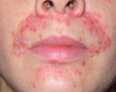
Aside from the area surrounding the mouth, perioral dermatitis lesions are also seen to affect other parts of the body. Lesions can also be seen around the eye (perioccular) and nostrils (nasolabial folds). Because lesions are not only contained around the mouth, it is now more correctly termed as periorificial dermatitis (peri-around, orifice-opening).
Perioral Dermatitis Image

Lesion on nasolabial folds

Who are the Ones Most Affected?

  • Children 6 months to 16 years old
  • Women 17-45 years old

Perioral Dermatitis: A Brief History

This condition sparks and interest among a few because this is a newly emerged disease. Considered to be a disease of the modern era, cases of perioral dermatitis have been recorded just in the recent years of the 50s and 60s. Interestingly, this is the same era when fluoride toothpastes and corticosteroids are used.

Perioral Dermatitis in Children

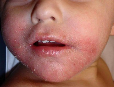
There are lesser incidences of perioral dermatitis in children. This condition is seen more common in adults than in people of younger ages. But nonetheless, there are still reported cases of perioral dermatitis among children. This condition affects children as young as 6 months to 16 or 18 years of age, and the condition can last up to 8 months.
Perioral Dermatitis in Children Image

Perioral Dermatitis in Children

Symptoms & signs in children

  • Papules, pustules or even vesicles appearing around the mouth
  • Same eruptions can also occur periorbitally (around the eyes) or perinasally (around the nose)
  • Eruptions are accompanied by pruritus
  • Eczematous in some cases
  • 70% of lesions are perioral
  • 43% occurs around the nose
  • 25% occurs around the eyes

Causes in Children

The exact cause of perioral dermatitis remains unknown, but steroid (whether topical or inhaled) is strongly related to the condition. Almost 72% of periorificial dermatitis in children is correlated with inhaled and topical corticosteroids. The remaining is also attributed to the use of:

  • Cosmetic agents
  • Fluorinated toothpastes
  • Topical fluorinated corticosteroids
  • Other irritants or chemical agents.

Signs and Symptoms

  • Emergence of small papules, pustules or vesicles around the mouth, nose or eyes
  • Erythema or redness in the areas of the lesions
  • Drying or scaling of the skin on the affected area.

Drying and scaling of the skin image

Drying and scaling of the skin

  • Itching or pruritus
  • Burning or stinging sensation (may occur after or during brushing)
  • Lesions forming a complete circle around the mouth except at the vermillion border

Causes

Corticosteroid Use

Perioral dermatitis is primarily attributed to corticosteroid use. Certain studies reveal that 71 out of 73 cases of perioral dermatitis have corticosteroid use. In children almost 72% used topical corticosteroids before the emergence of the condition.

The exact mechanism why topical corticosteroids cause such condition is still unknown. But experts speculate that topical corticosteroids reduce the protective skin layer and also decrease the size of skin cells (keratinocytes).

Indirect use of corticosteroids is also seen to cause or worsen perioral dermatitis; this is commonly seen in children who use inhaled corticosteroids to treat asthma. Long term use of these corticosteroid inhalers is also seen to have a relation with perioral dermatitis, even if it is not in direct contact with the skin.

Fluorinated Toothpastes/Creams

Another factor seen to cause perioral dermatitis is the use of fluorinated toothpastes, or other fluorine containing compounds. In a study conducted to 65 patients with perioral dermatitis, it was found that almost all of them used fluoride toothpastes. Replacing the toothpastes with non-fluoride relieved the patients of their symptoms.

Microorganisms

In certain cases, particular microbial specie is attributed to perioral dermatitis. Although studies are not yet conclusive, certain cases were seen to be related to the emergence of this condition. Microorganisms seen to cause perioral dermatitis include:

  • Fusiform Bacteria
  • Dermodexfolliculorum
  • Microscopic mites

Certain expert say that the “immune suppressing” effect of corticosteroids may have contributed to the proliferation of such microorganisms. This then leads to perioral dermatitis.

Use of Cosmetics, and other Facial Products

According to certain studies, the use of cosmetics and other facial products can also cause perioral dermatitis. Female patients using night creams, cosmetics, moisturizers and foundation predisposes them to developing perioral dermatitis by 13-fold (according to a clinical trial). Though the exact ingredient that causes this reaction is unknown, patients with perioral dermatitis are advised to discontinue the use of the offending product.

Treatment

Discontinuation of Topical Corticosteroids, Fluoride Containing Compounds, and Facial Products

Patients who have a history of topical corticosteroid use, fluoride containing compounds and other facial products are advised to discontinue the offending agents immediately. This would improve the patient’s condition and prevent further occurrence of the lesions.

Discontinuation of corticosteroid use can cause a short term flare of perioral dermatitis. To manage this, following interventions can be done:

  • Replacement with a less potent cream such as 1% Hydrocotisone or desonide cream
  • Topical calcineurin inhibitors
  • 1% Pimecrolimus to reduce the flares
  • Replacement of corticosteroids with Intramuscular injections

Oral antibiotics

Oral doses of Tetracycline 200-250 mg two times a day is highly effective in treating perioral dermatitis. Tetracycline treatment is usually given to adults or patients older than 12 years old. This is because the medication can cause enamel staining in children of a younger age.

Tetracycline Image

Oral Tetracycline Image

For children, 250 mg or erythromycin twice or thrice daily is seen to relieve the lesions and other symptoms of perioral dermatitis.

Minocycline and doxycycline 100 mg twice a day is also effective. For minocycline and doxycycline treatment should be as follows:

  • Full strength dose to last for 3-4 weeks (or until lesions are lessened)
  • Half strength dose to last for an additional 2-4 weeks until the rash has resolved
  • Topical Antibiotics

Application of topical antibiotics is seen to hasten the reduction of perioral dermatitis symptoms. Topical antibiotics such as the following can effectively treat perioral dermatitis

  • 1% metronidazole applied two times a day
  • 2% erythromycin ointment
  • Clindamycin gel

Diet for Perioral Dermatitis

Diet Rich In Vitamins A, E and B12

These vitamins promote a healthy skin. Vitamin A helps in regeneration new skin cells, while Vitamin E helps reduce inflammation and protects your skin cells from further damage. Vitamin B12, on the other hand strengthens the skin’s protective barrier preventing further damage and irritation.

Diet Rich In Vitamins A, E and B12 Image

Diet Rich In Vitamins A, E and B12

Failsafe Diet

The failsafe regimen stands for a diet free of additives, low in salicylates, amines and artificial ingredients. Such compounds can worsen or start an eruption, following this diet for at least 2 to four weeks can help lessen the lesions.

Probiotics and Vitamin C

Probiotics and Vitamin C share one action, and that is to boost the immune system in fighting infections and controlling inflammation. Since perioral dermatitis can be worsened by a secondary infection, preventing such would hasten your recovery.

Probiotics image

Probiotics must incorporate on patient with Perioral dermatitis diet

Is Perioral Dermatitis Contagious?

Perioral dermatitis is not a contagious disease. Contact with a person with perioral dermatitis doesn’t mean you would also acquire the same condition. Perioral dermatitis is an inflammatory skin condition; the primary cause of this is a person’s reaction to an offending agent.

The reaction is limited only to the person affected, the same agent that causes this cannot be spread to other people, and it may or may not cause the same reaction.


No responses yet

Anorchism

Apr 23 2016 Published by under Diseases & Conditions

Anorchism Definition

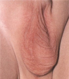
Anorchism is the medical condition where there is a unilateral or bilateral absence of the testes. This condition may be present as a congenital anomaly (present at birth) or due to secondary factors such as trauma or castration.

Anorchism image

Bilateral absence of testes or anorchism

What Happens When a Male Has Anorchism?

The testes serve as an important part of the male body, especially when it comes to the regulation of secondary sexual characteristics. The testes house the cells that produce androgens, and facilitate the production of sperm. With this organ absent, we can expect dysfunctions in the production or sperm and androgens

The absence of the testes can be classified into three general events:

  • Emergence of an ambiguous genitalia
  • Lack of secondary sexual characteristics
  • Impaired sperm production

Ambiguous Genitalia

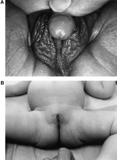
This happens when the testes did not develop while the fetus is in the womb. Sex organs begin to develop inside the womb at the eight week of pregnancy. Between the eight and tenth week, if the testes are lost the result is an ambiguous genitalia. This means in males, the penis appears smaller than the normal size. In some cases the reproductive organ appears to have both male and female attributes.

anorchia image


Ambiguous genitalia

Lack of secondary sexual characteristics

The testes produce male hormones called androgens. These hormones are responsible for making the male body distinct from the females. These distinctive attributes are called secondary sex characteristics. Secondary sex characteristics include growth of hair, deepening of the voice, broadening of the shoulder, development of the bones and muscles, and thickening of the skin.

In the absence of the testes, androgens would not be produced, and males would not achieve the secondary sex characteristics stated above. As a result males with anarchism appear smaller with less muscular built, there is absence of hair in the face, pubic area, arms and thighs, voice may sound child-like, and they are more likely to assume a body appearance that is thin and linear instead of the ectomorphic type with broader shoulders and narrow hips.

Impaired sperm production

The testis is also the structure which produces sperm cells. Within the testes is the structure called seminiferous tubules, this structure is responsible for sperm production. If the anorchia is bilateral, (both testes are absent), then sperm production is not entirely possible, but if it is unilateral (only one is absent) then sperm can still be produced.

Types of Anorchism

Anorchism can be classified according to the cause and presentation.

According to cause:

  • Primary anarchism – which can be attributed to defects present at birth or while the fetus is still inside the womb.
  • Secondary anarchism – loss of one or both of the testes as a result of castration, trauma, genial mutilation, infection, surgery or other underlying disease

According to presentation

  • Unilateral- only one testes is absent
  • Bilateral-both testes are absent

Anorchism Causes

Genes are seen to primarily cause anarchism, although genetics is a strong contender, the exact cause of the condition is yet to be known. Aside from genetics, other disease conditions are also seen to cause anorchism. These are the following:

  • Cryptorchidismundescended testicle Image

Undescended testicle as a cause of Anorchism

  • Vanishing testes syndrome
  • Swyer syndrome

Anorchism Symptoms

  • Both or one of the testes are absent
  • Failure to achieve puberty
  • No evidence of secondary sexually characters

Secondary sex characteristics are not evident which can be seen through the following:

  • lack of hair in the pubis, arms, legs, chest, and face
  • voice that remains to be child like
  • thin, non-muscular body
  • more fat accumulation than muscle
  • non-enlargement or growth of the penis
  • uneven waist to hip ratio
  • gynecomastia or breast development may be present

Anorchism Diagnosis

Anorchism can be diagnosed through the following procedures:

  • Physical examination– to diagnose the condition, the clinician would palpate the scrotum and feel the testes. The physical attributes of the patient is also examined. Here the clinician would look for the presence of secondary sex characteristics. When the patient does not exhibit these attributes, anarchism is suspected.
  • Hormonal tests – to further confirm if the patient has anorchism, hormonal tests are also done. Remember that the testis houses the cells that produce male hormones. Without the testes, production of the hormone is not possible. Anarchism is diagnosed when the levels of testosterone are low, and the level of Follicle stimulating hormone and luteinizing hormone are high.
  • Magnetic Resonance Imaging (MRI) and Ultrasound- these tests allows the clinician to view the scrotum for the presence or absence of the testes. MRI or ultrasound may show no gonadal tissues or in some cases the presence of just one. Imaging procedures such as MRI and ultrasound confirm the condition.

Anorchism Treatment

Hormonal Therapy

Males with anorchia are given lifelong supplements of androgens. Since there are no testes to produce the androgens, they are then supplied externally. With hormonal therapy, patients are expected to have improved conditions physically. This includes the development of the penis, development of the muscles and skeletal frame, and lesser fat deposition. Synthetic androgen hormones can be taken orally, or as a gel or cream that can be applied on the skin. Some of the synthetic hormones are given as a transdermal patch, or as an injection.

  1. Side effects of hormonal therapy – Hormonal supplementations also have certain side effects.
  • Oily skin and development of acne. This is because testosterone increases the activity of the sebaceous glands.
  • Increase in hematocrit levels. This side effect can be managed by regular venipuncture.
  • Worsening of sleep apnea and prostate cancer (pre-existing)
  • Thinning of hair

Testicular Prosthesis

The absence of testes is physically and psychologically traumatic for males. To aid this, testicular prosthesis is surgically inserted in the scrotum, to give revive the normal physical appearance of the male reproductive organ. In some cases, the prosthesis is also able to give hormonal supplementation much like what a real organ would do. Silicone implants are used as prosthesis. Since the treatment involves surgery, rigorous counseling of the effects and success of the treatment is done to the patient.

Complications of testicular prosthesis

  • Prothesis extrusion- this happens when the implant extrudes from the scrotum. This usually happens when the incision is made on the scrotum during insertion of the implant.
  • Silicone rupture
  • Disproportional or incorrect size or shaped prosthesis

Infertility Treatments

The major problem with anorchia is infertility. Since there are no testes, sperm production is not possible. Males may consider the following infertility treatments:

  • Intracytoplasmic sperm injection (ICSI)
  • Intrauterine Insemination of the sperm
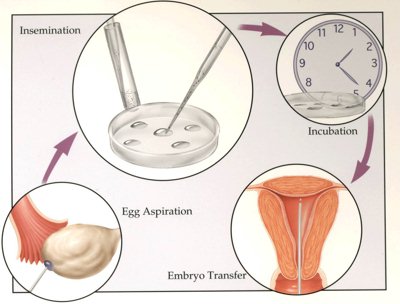
  • In Vitro Fertilizaion (IVF)

In Vitro Fertilization Image

In Vitro Fertilization to treat infertility brought by Anorchism

If the male cannot produce sperm at all (as in the case of bilateral anorchia), a donor sperm is used.

Complications of Male Infertility Treatments

The primary complication of male infertility treatments is its psychological impact on the couples. The process of infertility treatments can be tedious and success rates can be quite low. The process and the low success rates can be frustrating for both the male and female. This is why counseling and education on the procedure is done in order to prepare the couple psychologically.

No responses yet

Sesamoiditis

Apr 15 2016 Published by under Diseases & Conditions

What is Sesamoiditis?

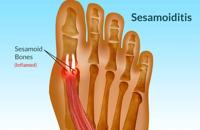
Sesamoiditis is an inflammation of the bones in the foot called the sesamoid bones and the tissues that surrounds it. Sesamoid bones are found below the large toe with an approximate size of a jelly bean. These bones are set in within a tendon and operate as protection and support to the adjacent tendon and improve its mechanical functions. Sesamoid bones are located in the knees, hands and foot. Sesamoiditis occurs mainly in people who are into running or dancing since it involves pushing off the toes. The sesamoid bones’ main function is to act like a pulley to increase the force of the tendons that controls the toe. And because of frequent pushing off of the toe, it becomes irritated resulting to sesamoiditis.

sesamoiditis

Characteristics of Sesamoiditis

  1. The swelling and inflammatory process usually occurs at the foot at the distal portion of the first metatarsal bone (big toe). It causes inflammation, pain, tenderness and swelling in the area.
  2. Periostitis or fresh bone growth can also take place along with sesamoiditis as manifested by an outward protuberance of the big toe

Once the sesamoids are inflamed and irritated, it is usually difficult to remove it because walking will always give strain on it.

Sesamoiditis Causes

Sesamoiditis can occur to any person at any age. Most of the time among younger individuals, this is due to trauma on the foot and stress.

Trauma

Sesamoiditis often results from excessive stress on the tendons and bones as a result of sudden flexion of the toes, wearing high-heeled footwear, and damage to the sesamoid bone. Excessive activity may also predispose a person for the occurrence of swelling in the sesamoid bones. Amongst women, those who wear high heels have greater chance of having sesamoiditis. This is because of the increased pressure on the forefoot. Athletes and dancers may also develop this problem over time.

Bony Foot

A bony foot usually puts added injury to the sesamoids because there are no enough subcutaneous layers in the soles of the feet to cushion the sesamoid bones.
Osteoarthritis

In older men and women, sesamoiditis occurs because of old age and the formation of added bones which lead to bone spur. The bone fragments can cause inflammation of the tissues that surrounds the area. Osteoarthritis is also another cause for sesamoiditis. Because of old age, the bones become weak which results to reduced ability in coping with the force exerted when walking.

Bone Deformities

Other causes include bone abnormalities wherein the sesamoid bones are larger than the jelly beans. The larger the bone, the greater force is to be exerted when walking which will increase the likelihood of injuries.

High Foot Arches

Also people with high arches have the tendency to have strained foot and overpronating can also result to sesamoiditis.

Sesamoiditis Symptoms

  1. Pain. Pain is the most universal symptom of sesamoiditis. This occurs mostly when walking, but even if simple weight bearing exercise can also cause pain. The pain is brought about by the tibial sesamoiditis since it is directly located at the base of the toe.

Tibial sesamoiditis is one of the common sesamoiditis wherein the medial or tibial sesamoid is affected. This is located at the inner edge of the foot and lies directly beneath the 1st metatarsal head.

  1. Gradual Onset. Sesamoiditis occurs gradually. The pain associated with sesamoiditis begins as mild and progresses to become severe.
  2. Swelling. Swelling is often seen in sesamoiditis. Bruising and redness may also be observed, but are more uncommon.
  3. Tenderness. Other symptoms include tenderness in the surrounding area and it worsens when there is pressure applied like walking or when the foot pronates.

Sesamoiditis Diagnosis

Sesamoiditis is easy to diagnose using the following procedures:

X-ray

Usually, with x-ray alone, it can confirm the problem. However, the sesamoids from time to time can be fractured and these do not usually become visibly clear on x-ray. In this case, a bone scan may be done to determine it.

Bone scan

A bone scan may also be ordered by doctors to correctly diagnose if it is a bipartite sesamoid or not.

Sesamoiditis Treatment

Sesamoiditis when it is not worse can be treated with home remedies to stop the pain and rest the foot. When these home remedies do not relieve the symptoms, it is always best to consult a physician.

Rest

Resting the feet is very important and then gradual activities may be added to avoid fatigue and pressure on the foot. Rest is the chief management to allow the foot to heal and reduce pressure on the area.

Sesamoiditis Taping

With the use or tape or athletic straps, sesamoiditis taping is done in order to immobilize the joint in the foot. This then allows healing of the sesamoid bone as well as the tissues and muscles surrounding the area. While the toe is being taped, it is important to avoid doing activities like running, dancing or even walking.

Ice Packs

Apart from taping the foot, it is also best to ice the sole of the feet. But it is not suggested putting ice directly since it will cause frostbite. Instead, use ice packs or wrap the ice with towel and apply it on the foot.

Shoe Modifications

If activities cannot be stopped such as walking, it is best to uses soft-soled and low-heeled shoe to decrease the pressure. Cushioning pads may be added too in order to feel comfortable.

Sesamoiditis Exercises

There are certain flexibility exercises that will help in stretching the plantar fascia, which is located below the arch of the foot. Sesamoiditis exercises can be done while on the process of recovery from the injury. While standing up, face the wall and then put a ball on top of the foot and the toes will be stretched upward. It is important to keep the heel on the floor. Then, stretch the knees facing the wall so that the arch of the foot will be stretched. Hold this position for 10 to 30 seconds depending on the person and the exercise can be repeated 3 times. This will help stretch the muscles of the foot and strengthen it in the process especially for those who are into running or dancing.

On the other hand, there are other forms of exercise alternatives such as swimming or biking. In that case, there is no need to pronate the foot which can result to sesamoiditis. Avoid more feet pounding exercises which will worsen the tenderness in the foot.

Sesamoiditis Surgery

If the Sesamoiditis is very worse, then it is time to consult the doctor. Especially with fractured bones, there is a need to perform operation in order to treat the problem and walk without pain. Sometimes if the fracture is worse, there is a need to remove the sesamoid bones, but it needs further assessment and planning since it can affect the toe and its function. A few surgical discomforts may be felt after Surgery like pain and swelling, but it will be better as days pass by. According to surgeons, there will be pain medications to be taken and using of crutches is useful to aid in mobilization. Full recovery may take months. But if there is no surgical incision, patients are expected to be walking, but with limited movements for 2-3 weeks. And for athletes who are into running, they need to wait about 2-3 more months to use their injured foot, but with limited range of motion.

Anti-inflammatory Drugs

Anti-inflammatory drugs are used to relieve pain and swelling. Common drugs administered are ibuprofen, aspirin and naproxen.

Sesamoiditis Prevention

Sesamoiditis is a mild foot condition that is highly preventable. Prevention of sesamoiditis is simple such as employing the following measures:

  1. Avoid using high-heeled shoes always. Use flat shoes from time to time.
  2. Apply shoe cushions.
  3. Engage in foot exercises after a day’s work.
  4. Rest the foot at night by elevating it in a pillow for 15 minutes to improve circulation to the bony prominences.

No responses yet

Red Bumps on Skin

Apr 07 2016 Published by under Diseases & Conditions

What are Red Bumps on Skin?

Bumps on the skin generally are abnormally raised areas of the skin. The cause of a bump goes a long way to indicate the type of bump. In the course of this article, we are going to discuss mainly the red bumps on the skin, and the following shall be taken to cognizance:

  • Red bumps on skin that itch

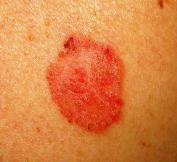
red bumps image

Image 1 – Red bumps on skin that itch

  • Red bumps on skin that do not itch
  • Small red bumps on skin

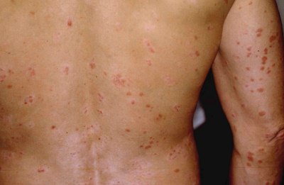
red bumps on skin


Image 2 – Small red bumps on skin

  • Raised red bumps on skin

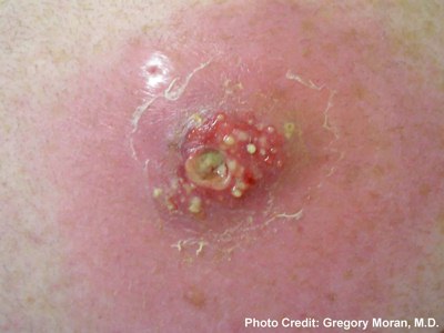
raised red bumps

Image 3 – Raised red bumps on skin

Furthermore, each of the above point can give a clue about the nature of the red bump. For instance, the first type – read bumps that itch – may indicate a bacterial infection, or bite from a heamatophagus insect (insects that sucks blood), or presence of a disease condition such as infection by the herpesvirus called “varicella-zoster virus”which cause chickenpox.

Red bumps on skin that do not itch may herald possible reaction to a chemical; an instance of which may be the release of histamines when you change your body cream and your dermal system is reacting negatively to the cream, possibly because of the presence of the presence of a substance to which you are allergic to.

Small red bumps on skin are usually allergic reactions, or mere erection of the arrector pili (the muscle attached to hair follicle which contract or relax to intrinsic and extrinsic environmental conditions) to from “goose bumps”.

Raised red bumps on skin may indicate mechanical injury.

As mentioned earlier, these do not confirm any of the instantiated conditions, unless by appropriate medical procedures.

Red Bumps on Skin Symptoms

In the course of our discussion, we made it clear that the presence of red bumps on skin does not always indicate the presence of a disease condition, or an infection. Therefore the type of the red bump will explicitly present its obvious symptoms.

In the case of red bumps on skin, there may be:

  • Slight increase in temperature, mostly in the case of red bumps on skin as a result of the presence of infective agents.
  • Cold
  • Irritation
  • Itching
  • Pain, especially in the case of infection

Red Bumps on Skin Causes

In a view to make this condition easily comprehended, red bumps are just normal dermal (skin) lesions that got filled-up with blood cell. Now, it does not just come straight like that; let us break it down.

Blood is the basic means of transporting or circulating vital substances that are needed throughout by the body cells. These circulated substances include: blood cells, oxygen, nutrients, enzymes, etc. In a view to bring this down to the understanding of all, basically when there is a presence of a foreign material in a particular area of the skin, skin receptors send message to the brain, and the brain try to “take the situation under control”. This may then require the brain to send command to some glands or organs to release some substances. If such substance released does not have a duct, it may have to be transported through the blood. Therefore, this can bring about the concentration of blood in the region – resulting in a typical red bump.

The scenario explained above explains common red bumps – which can also be the case during bruises, in which blood cells concentrates on the site of injury in order to prevent possible intrusion of germs or infectious agents.

The environments can also cause the occurrence of red bumps in some areas of the skin. This is evident in a situation whereby baby urinates in a napkin. If the napkin is made of an absorbent material and the wet napkin stays for a long time around its buttocks, small red bumps can develop on the skin in the region around the buttocks.

In the light of the information above, red bumps on skin is mainly caused by (but not limited to) the following:

  • Insect bite
  • Allergic reaction
  • Symptom of a disease
  • Mechanical injury
  • Presence of acne
  • Intrinsic and extrinsic environment

Red Bumps on Skin Diagnosis

Diagnosis entails the process of determining the nature of a disorder by considering the signs and symptoms exhibited by the patient and may also include medical observations.

Therefore, simple medical observation can diagnose the nature of red bumps on the skin. In cases of red bumps caused by infection, medical test may be required of diagnosis.

Furthermore, the environment in which the patient is can contribute a vital point to diagnosing the nature of the read bumps. Therefore, the diagnosis of red bumps on the skin can include:

  • Medical test
  • Physiotherapy: Especially in the case of red bumps on the skin that is influenced by the environment, whereby the environmental condition of the patient is countenanced.

Red Bumps on Skin Treatment

In the treatment of red bumps on the skin, treatment varies. Chemotherapy can be used. In this context, we say that chemotherapy is the treatment of red bumps on the skin by use of chemical substances. These chemical substances include pills and other solid, semi-solid or liquid chemical substances as such. In this case, mode of administration varies from topical to oral. In the case of oral administration, the patient takes medical prescriptions orally – through the mouth.

In the case of topical application, appropriate liquid or semi-solid chemical substances are rubbed locally on the affected region of the skin.

Materials that can be used to treat cases of red bumps on the skin include:

  • Topical application of Clotrimazole and Neomycin sulfate; especially in cases of acne caused by infection and other medications prescribed by your doctor for topical application
  • Oral administration of prescribed drugs
  • Administration of antihistamines like azatadine and acrivastine
  • Chimeraplasty – Chimeraplasty is an advanced method of treating red bumps. It is best applied to a situation in which red bumps are due to allergic reactions. It involves genetic tracing and manipulation in which the defective gene responsible for such allergic reaction is genetically engineered.

Red Bumps on Skin Remedies

There are various remedies by which the situation of someone with red bumps can be approached. One of such is ensuring a neat environmental condition, in which case, one avoids wearing wet clothes.

  • Improvising the environment as appropriate
  • Maintaining a serene and hygienic conditions
  • Proper used of prescribed medications

Prevention

In our discussion so far, you have been informed a lot about red bumps on the skin, and you will agree that it is not something that one will want to live with.

Therefore, it is imperative to discuss ways of preventing red bumps on the skin. In the light of this, the following practice can help in preventing red bumps on the skin:

  • Maintaining a hygienic environment
  • Consulting a professional or expert before using any body cream
  • Be conscious of the type of materials used

Complications

Although, red bumps on the skin are not a though not to crack, but if left unattended to, it can result in serious complications which can include the following:

  • In the case of red bumps on skin caused by infection, it can lead to weakening of the body immune system due to heavy invasion of the body system
  • If the above mentioned point prevails, it gives room for multiple infection, since the body immune system is no more so good
  • Disfiguring of the skin
  • Social implications – You will not readily want to sit beside someone with red bumps all over the skin

No responses yet

Acute Disseminated Encephalomyelitis

Apr 01 2016 Published by under Diseases & Conditions

What is Acute Disseminated Encephalomyelitis?

Acute Disseminated Encephalomyelitis resembles to the disease condition called MS or Multiple Sclerosis and its synchronism is ADEM. This kind of disease condition is a nonvasculitic kind of inflammatory condition which is considered to be as demyelinating. What happens is that instead of doing the function of the immune system which is to fight or get rid of the infection, the result is reversed where in it leads to inflammation in the person’s central nervous system which leads to demyelization. It has a characteristic of involvement of the brain’s white matter and spinal cord. Aside from that, it is known to be a monophasic kind of illness. It occurs in the association of either vaccination or immunization or perhaps the presence of systemic viral infection. Other experts would define it as a rare form of neurological disease condition. While others, consider it as an autoimmune and allergic for of encephalitis. And still others define it as a perivascular kind of inflammation that has a presence of edema. It is known that the children are mostly affected by this condition than adults. However, it can affect anybody.

Pictures of Acute Disseminated Encephalomyelitis

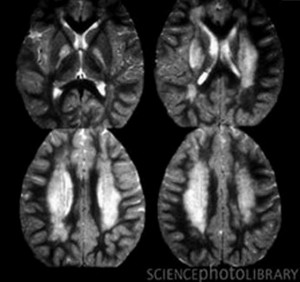
CT Scan indicating Acute Disseminated Encephalomyelitis

White Patches Resemble Inflammation in the Brain

Image Source: Science Photo Library

Acute Disseminated Encephalomyelitis Symptoms

Some people who are battling with this disease actually have infection first before acquiring ADEM. Children who have this condition often report to have severe and prolong aching of their heads. Aside from that, due to the presence of infection, inflammatory process is present in a form of fever. Aside from these, the persons manifest the following symptoms:

  • Dysphagia or difficulty in swallowing
  • Weak in their legs or arms
  • Falling and being unsteady
  • Confusion
  • Coma
  • Ataxia or unsteady in gait
  • Drowsiness
  • Fatigue
  • Vomiting
  • Seizure
  • Nausea
  • Upper respiratory tract infection
  • Visual disturbances such as optical neuritis
  • Muscular movements is another problem

Acute Disseminated Encephalomyelitis Causes

The exact occurrence of why ADEM occurs is unknown. But studies show that as mentioned earlier, a great deal of patients who are suffering from this disease actually had a history of infection prior to diagnosing them as having ADEM disease. The cause of which may be due to either:

Bacterial infection which includes:

    • Leptospira
    • Beta hemolytic Streptococci
    • Borrelia Burgdorferi
    • Mycoplasma Pneumoniae

Viral infection which may possibly include:

    • Measles
    • Mumps
    • Varicella zoster
    • Rubella
    • Enterovirus
    • Influenza virus
    • Epstein Barr virus
    • Herpes simplex virus
    • Hepatitis A
    • Coxsackievirus
    • Cytomegalovirus

Vaccination of either the following vaccines:

    • Varicella vaccine
    • Influenza vaccine
    • Polio vaccine
    • Pertussis vaccine
    • Hepatitis B vaccine
    • Rabies vaccine
    • Diphtheria vaccine
    • Measles vaccine
    • Rubella vaccine
    • Mumps vaccine
    • Pneumococcus vaccine
    • Japanese Encephalitis vaccine

Acute Disseminated Encephalomyelitis Treatment

The common and effective treatment of persons who are suffering from ADEM varies individually. What may be effective to one and may not be effective to others. The treatment is aimed at suppression of the aberrant immune systemic response. The common treatment includes:

Intravenous Immune Globulin therapy

This involves the antibody administration which is extracted from the person’s plasma. This is considered as the second choice. It is done with the use of blood product in order to reduce the possibility and risk of immune related disease.

Plasmapheresis

It is done through the use of machine which culls out the immune system component from the person’s circulation to be able to reduce the immune or inflammatory response. This kind of therapy takes for a few hours and is suggested to be done for 10-14 days maximum on alternative days. There is a need for central venous catheter to be able to rapidly remove the blood when necessary. Despite the benefits of this therapy, this therapy has its risk or bad effects such as discomfort, infections and bleeding tendencies.

Intravenous Corticosteroids

This is considered as the first line of drugs taken by patients who are diagnosed with such condition. This is suggested to be given for a week to be able to kill the bacteria or virus and will only taper the dosage after a week has lapsed. This medication is known for inflammatory reduction and will speed the recuperation process of the disease.

When one is on corticosteroids, the person needs to be monitored for low potassium level, disturbance in the sleep pattern, and blood glucose level. Aside from that, the side effect of this medication is change in one’s mood. Complications that are associated with this disease includes swelling of the face, flushing of the cheeks, gaining of weight, metallic like taste, and sleeping disturbance.

Chemotherapy

When the person is on the severe form of this disease, the physician may suggest that the person undergoes chemotherapy. Chemotherapy medication may be prescribed such as Mitoxantrone or Cyclophosphamide.

Acute Disseminated Encephalomyelitis Prognosis

Studies and statistically shows that there is approximately 10-30% of the mortality rate. Most often, children with this condition may have full recovery in a slow phase. And approximately up to 60-90% has been reported to have minimal to no deficits in the neurological aspect.

Patient who is under the treatment of corticosteroid or steroids, it can lead to the reduction of symptoms and prevent newer symptoms to develop as well as having a good prognosis. The prognosis for one person to another varies. The recuperation of one child will be slowly and will continue till it reaches full recovery with the help of treatment being provided. Some reports that they undergo recurring symptoms during their slow phase recovery. Such recurring symptoms include weakness, numbness or visual blurring. Aside from these, one must know that when a person is diagnosed with the severe form of this disease condition, there is a risk for fatality and some will be rediagnosed as having MS or Multiple Sclerosis. Also, when a person is diagnosed earlier with the disease condition and given the right treatment therapy without further delay, the overall prognosis will be good.

Acute Disseminated Encephalomyelitis Pictures

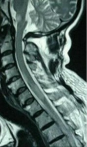
Acute Disseminated Encephalomyelitis pics

Demyelination of the Spinal Cord

No responses yet

© 2017 eHealthWall.com. All Rights Reserved. Privacy Policy
This website is for informational purposes only and Is not a substitute for medical advice, diagnosis or treatment.