Archive for June, 2012

Papilledema – Definition, Pictures, Causes, Symptoms, Treatment

Jun 28 2012 Published by under Diseases & Conditions

Papilledema Definition

Papilledema is the swelling of the optic disc caused by increased intracranial pressure. The swelling in the optic disc causes problems on the vision and may cause permanent vision loss when not managed. However, acute papilledema may result to preservation of vision.

The occurrence of papilledema usually develops over hours to weeks of increased intracranial pressure. The term Papilledema is only used correctly when it describes optic disc swelling as a result of increased intracranial pressure. Optic disc swelling with the presence of underlying infection and inflammation of the optic nerve is different from papilledema.

The optic nerve is anatomically continuous with the brain through the subarachnoid space. Any increase in pressure in the cranium thereby is being transmitted to the optic nerve. The anterior end of the optic nerve discontinues at the eye, thereby causing protrusion of the optic nerve on the area.

This results in the bulging of the ganglion cells of the optic disc. The prolonged and unmanaged edema of the optic nerve causes loss of the fibers of the ganglion cells and subsequently, impairment in the vision.

The incidence of papilledema is equal in men and women and may also occur at any age as long as the causes are present.
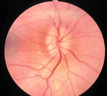
Optic disk edema image

Optic disk edema

Image source: webeye.ophth.uiowa.edu

Types of Papilledema

Bilateral Papilledema

Papilledema most commonly occur as bilateral, which means that both eyes are affected. Since the optic nerve is connected with the subarachnoid space, both sides of the optic nerve receive transmission of an increased intracranial pressure. The presence of bilateral papilledema usually rules out possible problems specific to the eyes that may cause swelling.

Unilateral Papilledema

Unilateral papilledema is a rare occurrence. Unilateral papilledema has only been documented few times. The development of disc swelling in only one eye implicates the occurrence of other etiologic phenomenon.

Grading of Papilledema

  • Stage 0 – This stage describes normal pressure in the optic disc.
  • Stage 1 – This involves very early papilledema where there is obscuration of the disc in the nasal border.
  • Stage 2 – This stage involves the presence of obscurations of all borders with elevation of nasal border. This is described as early papilledema.
  • Stage 3 – There is already increased optic nerve head diameter and considered moderate papilledema.
  • Stage 4 – This is marked papilledema involving total elevation of the nerve head with peripapillary halo and total obscurations.
  • Stage 5 – Severe papilledema involves obliteration of the optic cup and anterior expansion of the head of the optic nerve representing severe damage on the nerve.

Papilledema Symptoms

Papilledema is usually asymptomatic during the early stage of the condition. When symptoms occur, they include:

Headache

Headache occurs as a result of the increased intracranial pressure. Headache is usually an early sign of increased intracranial pressure and a developing papilledema. The nerves become compressed as a result of high pressure leading to severe headache. Headache usually becomes severe upon awakening and become worsened by Valsalva maneuver such as coughing and straining on stools.

Nausea and Vomiting

The increased intracranial pressure also puts pressure in the vomiting center of the brain, which is the medulla oblongata. The characteristic vomiting of increased ICP is projectile vomiting.

Blurring of Vision

The swelling of the optic disc causes the disturbance in the vision because the nerves are responsible for the sensory function of the eyes.

Visual Obscurations

This involves inability to see in certain areas of the visual field as a result of optic disc swelling also termed as blind spots.

Loss of vision

When the swelling becomes persistent, the optic nerve becomes permanently damaged leading to visual loss. The occurrence of visual loss happens during the last stage of the condition.

The eyes are examined using an opthalmoscope and may reveal loss of venous pulsation, venous engorgement, hemorrhages, optic disc elevation and radial retinal lines termed as Paton’s lines.

Papilledema Causes

Causes of papilledema include all factors that may increase the intracranial pressure such as:

  • Space occupying lesions or tumors in the CNS – The cranium and the vertebrae is a closed system, which means that any foreign body or tumor in the area causes the pressure to increase leading to increased intracranial pressure.
  • Decreased CSF resorption – The CSF is continuously circulated in the ventricles in the CNS. The CSF is also reabsorbed to make way for new CSF to be circulated. However, in the cases of inflammation in the meninges or venous sinus thrombosis, the CSF is poorly reabsorbed thereby increasing the circulating CSF and pressure inside the cranium.
  • Idiopathic Intracranial Hypertension – This is an increase in the pressure in the arteries inside the cranium leading to increased pressure inside the cranium.
  • Cerebral edema – Cerebral edema is a result of various medical conditions. It is commonly caused by trauma in infection. The edema or swelling increases the pressure and thereby leading to papilledema.
  • Medications – Medications such as corticosteroids, minocycline, Accutane, lithium and nalidixic acid causes increased intracranial pressure during drug withdrawal and overdose.

Papilledema Diagnosis

The diagnosis of papilledema relies on the use of slit lamp examination and opthalmoscopy showing a characteristic of the optic nerve disc. The diagnostic tests are done when new onset headache is noted with possible increased intracranial pressure. Aside from the optic nerve diagnosis, a biopsy can be made to detect whether the tumor is benign or malignant whenever it is present.
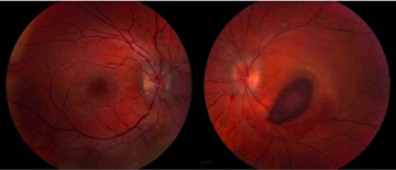
papilledema image

Papilledema Image

Differential Diagnosis

Papilledema is usually differentiated from other conditions involving the eyes such as papillitis, optic neuritis, glaucoma, and retinal detachment.

Papilledema Treatment

The treatment of papilledema is primarily focused on the treatment of the underlying cause of the increased intracranial pressure. The direct cause, which is increased intracranial pressure, can be managed using the following treatments:

Diuretic Administration

The use of diuretics such as lasix and acetazolamide increases the adsorption of CSF to be eliminated through the urine.

Steroids

Steroids are potent anti-inflammatory medications and may be given for patients with cerebral edema. However, steroid use should be monitored because any abrupt change in the dosage may also cause increased ICP.

Surgical Approach

Repeated lumbar punctures are usually done to remove excess CSF in the spine thereby reducing the overall circulating CSF in the brain.

Lumbar Puncture Image

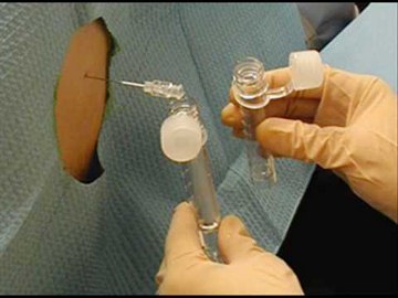
Lumbar puncture to remove excess CSF

Medication Modifciation

Medications causing the increased ICP should be stopped to prevent further increase in pressure.

Papillitis Vs. Papilledema

Papillitis is a condition of optic neuritis. It is called intraocular optic neuritis wherein there is inflammation of the optic nerve head. Papilledema on the other hand, is the bulging or swelling of the optic disc. Papillitis involves significant loss in the visual field and pain on the globe of the eyes, which is not present in papilledema.

No responses yet

Bleeding After Menopause Causes, Diagnosis, Treatment, Complications

Jun 24 2012 Published by under Diseases & Conditions

What is Vaginal Bleeding after Menopause?

Bleeding after Menopause is also called as Post-Menopausal Bleeding (PMB). After reaching your menopausal stage, in rare cases you begin to bleed again. It may be confusing in a woman’s point of view and somehow frightening because of its occurrence.

If a woman already reached her menopausal stage, it is expected that the regular menstruation will stop. But in some cases, bleeding after menopause occurs due to certain underlying factors.

Bleeding after Menopause

Characteristics of Bleeding after Menopause

  • It is considered post-menopausal bleeding if a woman has gone a year with no period and then has it again.
  • Post-menopausal bleeding may occur as heavy flow or light flow.
  • Women experience this kind of condition almost 20-30% of their population.
  • It is common to have post-menopausal bleeding that is due to hormonal shift; the uncommon scene of post-menopausal bleeding is that there may be abnormality in the cells within the uterus.
  • Along with these factors, this can be a symptom of hyperplasia of the uterus a minor abnormality in a woman’s body, or of a severe condition; a Uterine cancer.

Uterine Cancer

Bleeding after Menopause as a symptom of uterine cancer

All of these conditions are considered highly treatable, but it is the woman who initiates to have this checked to be treated as soon as possible.

In the case that you have bleeding after a long period of time after menopausal, it is best to consult a physician.

Associated Symptoms of Bleeding after Menopause

These are common symptoms you might encounter along with vaginal bleeding after menopause:

  • Cramping or Abdominal Pain
  • Abdominal, pelvic, or lower back pain that can be severe
  • Severe pain on the abdominal, pelvic or lower back part
  • Pain in sexual intercourse duration
  • Fatigue

Other symptoms that occur along with vaginal bleeding after menopause include:

  • Change in bowel movements
  • Signs of Constipation
  • Irritability and mood swings
  • Rapid heart rate (tachycardia) or fast heart rate as a result of hypovolemia
  • Sudden weight gain or loss
  • Fever and chills with ill feeling associated with infection

These symptoms indicate presence of further problems in a woman and these should not be ignored. Early detection of underlying causes is important to immediately implement certain treatments.

What Causes Bleeding After Menopause?

Bleeding after menopause can be caused by the following factors:

  • Trauma – Trauma can result from sexual intercourse or any foreign body.
  • Poor vaginal lubrication – Menopausal women have decrease in vaginal lubrication because of hormonal changes. Because of this, sexual intercourse may be painful or may result in bleeding.
  • Hormonal Replacement therapy – Hormonal replacement therapy may cause vaginal bleeding because of fluctuations in the estrogen and progesterone levels.
  • Steroids and Anti-coagulants – These can also cause post-menopausal bleeding because of impaired clotting mechanism of the blood.
  • Cancer medications -Treatments for cancer, specifically breast cancer may result in post-menopausal bleeding as a side-effect
  • Clotting problems as a result of kidney, thyroid or liver disease – These conditions often lead to bleeding because of impaired synthesis of clotting factors.
  • Infections – Infections in the reproductive tract such as Chlamydia can also cause post-menopausal bleeding.
  • Malignancies – Cancer in the reproductive tract also leads to post-menopausal bleeding.
  • Nutritional state, diet, exercise and stress
  • Endometriosis – Endometriosis is an excessive proliferation of the lining of the uterus leading to dysfunctional bleeding.

endometriosis

Endometriosis as a cause of bleeding after menopause

How to Diagnose Bleeding After Menopause?

If you have a bruise or notice to bleed so easily, consult a physician because this may mean a bleeding disorder.

Physicians usually diagnose post-menopausal bleeding based on the following tests:

Complete Blood Count

This is done to check the RBC and platelet component of the blood. Low platelets tend to cause bleeding because of reduced platelet aggregation (a step in the clotting process). The RBC, hemoglobin and hematocrit level are also checked to determine extent of bleeding because it may cause anemia.

Partial thromboplastin time (PTT), prothrombin time (PT) and bleeding time determination

These are also checked to evaluate the clotting mechanism in the body. An elevated result may cause bleeding in all areas in the body, including vaginal bleeding.

Thyroid evaluation

Thyroid hormones are also checked because a decrease in thyroid hormones leads to vaginal bleeding.

Pap smear

Pap smear is done to detect cervical cancer.

Pelvic Exam

This is usually done by gynecologist to examine the pelvic organs. Also there are more specific tests that may detect what causes vaginal bleeding after menopause. These include:

  • Transvaginal ultrasonography (TVUS) – This kind of test can look closely at the organs in the pelvic area. PMB ultrasound is able to show the lining and thickness of the womb. Thus, this can show whether the patient has chances of getting cancer or not.
  • Pipelle test – This test is performed along with the TVUS to examine the endometrial cells.
  • Out-patient hysteroscopy. If the TVUS and the pipelle test have limited results, the hysteroscopy can be done as an out-patient basis. This test uses a thin scope inserted through the cervix and into the uterus to visualize the area.
  • Biopsy. It is a test that involves getting a small sample of the endometrial lining for histologic analysis. This could also be carried out as an out-patient basis.

endometrial biopsy for bleeding after menopause

Endometrial Biopsy to determine the cause of bledding after menopause

How to Treat Bleeding After Menopause?

There are different kinds of treatment for vaginal bleeding after menopausal, but it is most recommended to see your doctor first for accurate treatment and diagnosis. Managements applied to manage vaginal bleeding problems include:

Dilatation and Curettage (D&C)

This procedure is done under general anesthesia. It involves scraping the uterine lining to remove dysfunctional cells that lead to dysfunctional bleeding.

dilatation and curettage

Dilatation and Curettage to treat bleeding after menopause

Platelet transfusion

When platelet counts are low, platelet transfusion is started immediately to prevent profuse bleeding.

Anti-fibrinolyitc therapy

Medications such as Tranexamic acid is given to prevent dissolution of fibrin clots that leads to bleeding. However, anti-fibrinolytic therapy should be carefully used in patients with history of stoke because it promotes blood clots that may be a reason for cerebrovascular accidents.

Remedies for PMB

  • Avoid using tampons that can irritate the lining of the vagina. Use pads instead.
  • Make sure to change pads every two hours whether it is not soaked. Presence of old blood around the vagina may cause bacterial growth and may lead to sepsis.
  • Avoid using anti-coagulants and aspirins to prevent bleeding as a side-effect.

Complications of Bleeding After Menopause

Post-menopausal bleeding needs prompt consultation from a doctor. The post-menopausal bleeding or PMB may be a sign that you have certain conditions that should be attended by health professionals. Complications of such could lead to more severe cases such as:

  1. Sepsis – Prolonged bleeding encourages bacterial growth that may spread to the blood and other areas of the body.
  2. Hypovolemia – Blood volume depletion may also result from prolonged bleeding.
  3. Shock – This may be due to septic shock or hypovolemic shock as manifested by severe hypotension and circulatory collapse.

The presence of vaginal bleeding after menopause should warrant the individual to seek medical attention because complications are often life-threatening. Minor forms of vaginal bleeding such as in the case of trauma, medication side-effects and hormone replacement therapy should still be evaluated to detect any serious underlying condition.

No responses yet

Cherry Angioma – Pictures, Removal, Causes, Home Treatment

Jun 18 2012 Published by under Diseases & Conditions

What is Cherry Angioma?

Cherry angiomas are cherry-red eruptions on the skin as a result of proliferation or dilation of blood vessels in the area. Cherry angiomas are the most common kind of angiomas. They are also known as Campbell De Morgan spots owing to the one who discovered it during the nineteenth century, a British surgeon named Campbell De Morgan. These cherry-red papules are also termed as senile angiomas because they occur at old age. They are benign and do not result to malignancies. It may occur on almost all areas of the body, but the most common site are the face and torso.

What causes cherry angioma?

Cherry angiomas usually develop during middle age, but it can occur in younger people. The main cause of the development is rarely understood because people’s interest has not been on it since it does not cause malignancy. A recent study indicates that a defect in the RNA causes the blood vessels and capillaries to proliferate. Specifically, a reduction in microRNA 424 levels is seen in patients with cherry angiomas. This causes increased protein expression and subsequently endothelial proliferation giving rise to dilation of capillaries on the skin. Aside from this, certain risk factors include exposure to chemicals such as butoxyethanol, mustard gas, bromide and cyclosporins. Genetic inheritance also spreads the disease to other people. Stress has also played a role in the formation of cherry angiomas. Emotional, physical and psychological stress leads to faster aging. Development of cherry angiomas increases with age. Cherry angiomas also occur in all races and in both sexes.

What are symptoms of cherry angioma?

Cherry angiomas are made up of groups of dilated capillaries that are evident on the skin. Signs and symptoms include:

  • Cherry-red to purple bumps
  • Present in the torso, hands, arms, legs, face, scalp and neck
  • Young angiomas are the size of a pinhead, but it grows to about .25 inch in diameter
  • Spongy, smooth and mushroom shaped
  • Painless and harmless
  • Bleeding occurs when injured. Cherry angioma on the scalp usually bleeds due to accidental brushing or combing of the hair.

How to Diagnose Cherry Angiom?

Cherry angioma is usually diagnosed with the physical examination of the bump and studying the characteristics. Further tests are not required, but a biopsy may be done to confirm the diagnosis and also helps in the removal of the angioma.

Cherry angioma treatment

Cherry angiomas do not require removal as these are not malignant and they are harmless. Treatment is only considered when they cause frequent hemorrhage or irritation. However, people with cherry angiomas may prefer removal for aesthetic purposes. Minor surgeries are done and medical treatment is not indicated because they do not cause a certain disease and are benign. Patients need to ask their doctors several questions before undergoing treatment to explore the benefits and risks as well as alternatives. Patients may ask:

  • What are the methods of removal available?
  • Is scar formation possible after treatment?
  • What are the risks of undergoing angioma removal?
  • Are there any alternatives for surgery?
  • Is cherry angioma preventable?
  • Does exposure to ultraviolet rays affect the angioma?
  • Will angiomas lead to carcinomas or cancer?
  • How do we prevent occurrence of cherry angiomas?

Adequate knowledge regarding the angioma may help in decision making and planning for interventions. The following therapies are done for cherry angioma removal:

Electrosurgery or Cautery

Electrosurgery is done by dissecting the angioma using cautery, a special electrical instrument likened to a needle. Scarring is minimal when expert operators are the ones who perform the procedure. Bleeding is also prevented because of rapid closing of bleeders. Local anesthetic is applied prior to the cautery.

Laser Removal

Pulsed dye Laser – Cherry angioma laser removal involves the use of an organic dye mixed with a solvent as the lasing medium. Absorption of the laser energy is possible in the hemoglobin, but this does not cause significant effects.

Intense Pulsed Light (IPL) – IPL is a more advanced method of laser surgery involving more concentrated beam light.

Shave Excision

This involves the delicate removal or slicing of the angioma using a blade in a horizontal angle. Specimen is subjected to biopsy to confirm the diagnosis. Bleeding is prevented through cautery of blood vessels.

Cryotherapy

Cryotherapy or cryosurgery involves the removal of the cherry angioma through freezing by the use of extremely cold substances such as liquid nitrogen (nitrous oxide), carbon dioxide, argon, and dimethyl ether. It also involves destruction, irritation and coagulation. It is a minimally invasive surgery and may result to mild pain that can be reduced by the use of analgesics.

Curettage

Performing curettage is not commonly used like the more conservative therapies discussed earlier. Curettage is the scraping of the angioma using a curette (an instrument used for scraping). Never remove angiomas on your own to prevent profuse bleeding, scarring and infection. Doctors are the ones trained to remove a cherry angioma to arrive with best results.

Natural Remedies for Cherry Angioma

Natural remedies for cherry angiomas are more favored because they are less invasive and less expensive. However, effects may not be seen immediately. Natural remedies are more suited as prevention for cherry angioma formation. The following treatments are the suggested home remedies for cherry angiomas:

Diet modification

Include more fruits and vegetables because these help in increasing the texture and elasticity of the skin. Better skin outcomes mean lesser development of cherry angiomas. Avoid foods that are processed such as canned and junk foods. Include Vitamin A and Vitamin D supplements to promote young and healthy skin.

Topical herbs

Apply sandalwood and basil leaves cream on the cherry angioma to reduce the appearance. These can be pounded and the juice applied on the skin. Witch hazel also reduces bacterial growth, preventing angioma formation

Apple cider vinegar

Traditional remedy includes applying apple cider vinegar to the angioma to dry up the bump. Some individuals who have tries said that the angioma fell off after two weeks of application.

Drink plenty of fluids

Increase water intake to 1.5 to two liters per day. Consume more fluids in the form of juices, jellies and shakes. Good skin hydration prevents angioma and skin breakdown because it enhances the moisture and elasticity.

Avoid stress

Stress is a major risk factor for cherry angioma formation. Learn to do stress reduction activities such as focused breathing, yoga, guided imagery and listening to music. Prevention of stress means prevention of cherry angioma.

Cherry Angioma Prognosis

These cherry-red bumps are most often benign and harmless. It increases in size and number with age, but a sudden development of several angiomas may signal a developing internal malignancy.

Cherry Angioma Complications

Possible complications of angioma include:

  • Bleeding when injured, but will easily stop upon pressure application and does not lead to profuse bleeding
  • Psychological distress as a result of body image disturbance. These people usually opt to removal of the angioma for aesthetic purposes.

Cherry Angioma Pictures

cherry angioma

Cherry Angioma pictures

Cherry Angioma images

 Cherry Angioma pics

Cherry Angioma

Cherry Angioma photos

Cherry Angioma on skin

Cherry Angioma on Face

The most common site of blood vessel proliferation is in the face. The vascular lesions are characterized as cherry red to purple in color and flat topped. They usually grow as the individual increase in age and may increase in about few centimeters in size. Some angiomas are close to each other and form a polypoid angioma or a multiple adjoining angioma. These may seem larger and more apparent. Cherry angiomas on the face are sensitive and may cause bleeding when the area is injured. Cherry angiomas on the face also lead to body image disturbance because of the red eruptions on the skin. Although the vascular lesions are harmless, occurrence on the face may result in loss of self-esteem. Removal of the angioma in these people may be considered.

Cherry Angioma on face

No responses yet

Onychomycosis (Nail Fungus) – Treatment, Pictures, Sub types, Causes

Jun 13 2012 Published by under Diseases & Conditions

What is Onychomycosis?

Onychomycosis is a disease condition that affects the person’s nail plate found in either the toes or the fingers. It is a kind of fungal infection. Most often, the reported cases are the toenails. Some experts would call it as tinea unguium, which is known as one of the largest subclass of this nail disorder. When left untreated, it leads to progressive and insidious destruction of the person’s affected nail plate. It is most commonly reported in males in the elderly ages around 40 to 80 years old.

Subtypes of Onychomycosis

There are actually three common subtypes of Onychomycosis, namely:

1. Proximal Subungual Onychomycosis (PSO)

It is a kind of fungal infection that penetrates in the proximal part of the nail fold. Amongst the three subtypes, this kind of subtype is the least common especially in people who are healthy. It is more commonly reported in persons who are known to be immunocompromised or who have an immune system that is impaired. Its etiological factors are trichophyton rubrum and fungus.

What happens here is that the fungus will enter the person’s nail bed by the proximal part of the nail fold and the skin flap that covers the nail which is located behind the affected cuticle. The root, which is the place where the nail is made, is where the fungus forays and then right after that, it will precede towards the nail’s edge.

2. Distal Subungual Onychomycosis (DSO)

Amongst the three common subtypes of Onychomycosis, this is reportedly to be the most common subtype. The etiological factors that are associated with this kind of nail disorder are dermatophytes, fungus, and trichophyton rubrum.

It begins under the person’s nail which is located at the affected finger or toes’ edge. Just like the proximal Subungual Onychomycosis, the fungus will eventually spread over the matrix of the nail and will later on inflict both the nail bed and the nail itself. The person who has this kind of subtype will experience nail plate detachment from his or her nail bed as well as discoloration on the nail plate which is affected. In addition to that, his or her nails become thicker than before. Aside from that, persons with this nail disorder subtype will report to feel uncomfortable and painful especially whenever they wear foot wears or shoes, for that matter.

3. White Superficial Onychomycosis (WSO)

This last and final common subtype is the known to be the second commonly reported subtype of infection of the nails. The causative agent associated with this subtype is trichophyton mentagrophytes and dermatophytes.

When a person has this kind of subtype, he or she will manifest white patches which are tiny and appearing on the nail plate surface. When the fungal infection spreads, the affected nails will become dry out and eventually the nail’s surface crumbles and flakes. According to studies, it becomes like that because the fungus will eat on the nail keratin and protein for its nourishment leading to the weakening of the nail plate and eventually distorting it.

There are actually three kind of white superficial Onychomycosis or WSO which are dual and classical nail plate invasion, the ventral and superficial kind and lastly the pseudo kind of WSO that have fungal invasion in the affected nail plate, which is deep.

Onychomycosis Image

Nail changes as one of the symptoms of Onchomycosis

Onychomycosis Symptoms

Persons, who are diagnosed with this kind of nail disorder, will manifest the following symptoms associated with this kind of nail disorder, which are as follows:

  • Nail shape changes
  • Nail having debris trapped under it
  • Loosening of the affected nail
  • Thickening of the affected nail or medically termed as hyperkeratosis
  • Discoloration or leukonychia such as yellow or white streaks on the nail’s side
  • Lost in the shine and luster of the affected nail
  • Brittleness
  • Edges of the affected nail crumbles
  • Paresthesia
  • Opaque or cloudy appearance of the affected nail
  • Inflamed affected nail
  • Onycholysis or when there is a separation of the nail plate from the nail bed
  • Losing of dexterity
  • Discomfort
  • Pain

According to studies, as the disease will progress, there is a big possibility that it will interfere with the person’s ability to walk, stand and do exercises.

Onychomycosis Causes

The etiological reason or factors or pathogens that are associated with the nail disorder called Onychomycosis are believed to be the following:

  • Candida – It is an etiological factor or pathogen that is frequently reported to be involved in the subtropical and tropical region which is known to have climates that are humid and hot.
  • Nondermatophytic molds – Just like Candida, the Nondermatophytic molds are frequently reported to be seen in patients who lived in the tropical and subtropical regions where their climates are humid and hot.
  • Dermatophytes – The dermatophytes are what are known to be the fungi which are most responsible for this kind of nail disorder especially when the person who was inflicted in the disorder lives in the western countries.

Onychomycosis Treatment

With regards to treating Onychomycosis, experts say that it will be challenging to treat such kind of nail disorder which is brought about infection that is known to embed within the affected nail that is difficult for anybody to reach. The expected removal of the symptoms that the patient manifest will be actually slow and have the possibility to go through the recuperation phase for a year or more than that, depending on the respond of the patient to the treatment given. The common treatment which is suggested and is expected under this kind of nail disorder includes:

Pharmacological Treatment

Systemic and topical treatments are prescribed. Studies show that combination of both medications and treatments will increase the rate for recuperation of the patient. Most often the classification of the drug or medication prescribed are those belonging in the antifungal classification.

Surgical Treatment

This is the last treatment option for this kind of disease condition or nail disorder. The kind of surgical procedure done will be different per patient and will greatly depend on the physician’s assessment.

Onychomycosis Pix

Surgical removal of the affected nail

Laser Treatment

This kind of treatment represents future option for treatment. Still, further studies are conducted. Although, some podiatrist have tried this kind of treatment to patients with Onychomycosis yet the efficacy as a treatment is still debatable at this point.

Alternative Treatment

Some would prefer alternative medicine such as grapefruit seed extract, snakeroot leaf extract, and Australian tea tree oil to cure Onychomycosis.

Onychomycosis Pictures

Onychomycosis pictures

Image credit – skinsight.com

Onychomycosis images

Source – myfootshop.com

Onychomycosis photo

Onychomycosis (fungal infection)

Onychomycosis pics

No responses yet

© 2017 eHealthWall.com. All Rights Reserved. Privacy Policy
This website is for informational purposes only and Is not a substitute for medical advice, diagnosis or treatment.