Archive for March, 2012

Retroperitoneal Fibrosis – Causes, Symptoms, Treatment and Diagnosis

Mar 12 2012 Published by under Diseases & Conditions

What is Retroperitoneal Fibrosis?

Retroperitoneal Fibrosis also called as Ormond’s disease named after John Kelso Ormond who rediscovered the condition in 1948. Retroperitoneal fibrosis commonly happens within the fibrous tissue; specifically in the retroperitoneum and in the internal organs of the body (eg. Kidneys, aorta, renal tract). Symptoms of such are deep vein thrombosis, renal failure, hypertension and the most common is the lower back pain. Damage done in the kidney may be there temporarily or permanently if not given attention to or not been aided quickly.

Also, the idiopathic retroperitoneal fibrosis exists and is a part of the disease inclusion with the “Chronic periaortitis” that may include the inflammation of the abdominal aortic aneurysms and perianeurysmal retroperitoneal fibrosis. By not studying it further it is similar, further more the periaortic fibrous tissue in the perianeurysmal fibrosis and may cover structures and cause complications; in cases with inflammatory aneurysms this does not occur.

Retroperitoneal Fibrosis Symptoms

Early symptoms

  1. A pain in the abdominal area that may increase its tension in time
  2. Change of color in the legs due to decreased flow of blood
  3. Pain in legs due to decreased flow of blood
  4. Swelling of the leg

Later symptoms

  1. Urine output has decreased
  2. No sign of urination (anuria)
  3. Vomiting, Nausea and affected thinking that is also caused by kidney failure; also with toxic chemicals in the blood
  4. Uncontrollable pain in the abdominal area with hemorrhage due to death of tissue in the intestines

Thus, these symptoms and signs that is associated with retroperitoneal fibrosis are not specific. Diagnosis of such disease may require a visit to your physician.

Causes

The Retroperitoneal fibrosis is not a common disorder among people by which the ureters or tubes that function as the passage of your urine from the kidneys to the bladder is massively blocked behind the area of the stomach and intestines. The ratio of three is to one of the cases are prone to malignancy of the mass. Physicians don’t have the idea why this masses come up to this form. This disease is commonly found in men than in women and most likely found between the ranges of ages 40-60.

Diagnosis

Getting basis on the result of the laboratory studies it is expected that the retroperitoneal fibrosis cannot rely to its diagnosis as accurate results, as it is not familiar with the disease and cannot be applied accurately by an individual. In our technology today, CT scan is the best modality that can detect the said disease. On the other hand, biopsy is an option though it is not recommended for the fact that it is proper when malignancy or infection occurs. The biopsy test should also be applied to the patient who does not respond to initial treatment given or if the location of the fibrosis is not in a common position.

ct scan in retroperitoneal fibrosis (Ormond’s disease)

Abdominal CT scan showing Thickening in retroperitoneum.

There are some other tests also available that may be able to detect this condition:

  1. Intravenous pvelogram (IVP)
  2. Kidney Ultrasound
  3. MRI of the abdomen
  4. BUN and creatinine blood tests

Treatment

Upon treatment of the retroperitoneal fibrosis doctors include:

Corticosteroids

Powerful anti-inflammatory medicines that help regulate inflammation. Corticosteroids are also used supportively to prevent nausea. Also preventing nausea as one of the symptoms of retroperitoneal fibrosis.

Tamoxifen

Tamoxifen is a drug given to block the effects of the hormone estrogen. There are common reports that say tamoxifen is very useful on treating retroperitoneal fibrosis. Also, this is to improve the condition in various small trials.

Biopsy

A biopsy should be done to confirm the diagnosis of the retroperitoneal fibrosis. This can be the key to diagnose the alterations of renal function.

Surgery Stents (Draining Tube)

This is the procedure of draining pus, blood or other body fluids from a wound. These are performed by surgeons or interventional radiologists. The drain inserted in the wound not aim for faster healing of the wound but to drain body fluid which might be the focus of infection and by which it can be prevented.

Immunosuppresive Therapy

There are alternatives you might be able to try to treat retroperitoneal fibrosis. The main goal of the therapy that is achievable are to create a relief the obstruction that is caused by fibrosis, stop the progress of the fobrotic process and prevent the occurrence of the disease. If it is cause by another factor, treatmant might be aimed by the feature and factors of etiology. Initiating immunosuppresive therapy is common if the disease is idiopathic.

In most cases that involve the aorta can step up the surgical process to removal of the mass and can be extremely dangerous that is near impossible to succeed with this kind of operation. Idiopathic malignancy should be excluded in precipitating causes along with most cases. While surgical ureterolysis has been the preferred primary option of treatment, because it can allow the biopsy specimens to be kept while urethral blockage has been stopped or relieved.

Managing the retroperitoneal fibrosis includes:

  1. Suppressing inflammatory processes
  2. Reducing the morbidity
  3. Preserving renal function

Thus, avoid lone-term use of such medications that have methysergide, which can cause retroperitoneal fibrosis. The methysergide is sometimes used to treat headaches such as migraine.

Complications

Possible complications of retroperitoneal fibrosis may lead to cases like:

Chronic Bilateral Obstructive Uropathy

It is the long-term blockage with the flow of your urine from both of your kidneys. It is a slow blockage that may get worse over time. This may be caused by bladder outlet obstruction. In this case, the kidneys normally produces urine but the urine passage is blocked. This is why the urine cannot leave the bladder, as these happens the urine backs up and causes the kidney to swell. It is most commonly found in men that may result to the enlargement of the prostate, also known as benign prostatic hyperlasia (BPH).

Other factors that cause these complications:

  1. Urethral stones that are bilateral
  2. Tumors on the bladder
  3. Tumors on the prostate
  4. Other structures around the uterus, bladder neck or urethra, which may be tumors or masses
  5. Tumor or retroperitoneal fibrosis
  6. A scar tissue or birth defect that may cause the urethra to narrow
  7. Neurogenic bladder

Chronic Kidney Failure

It is slowly loosing the kidneys’ function over time. It is the loss of function of the renal over time. Reduced appetite and feeling unwell are the symptoms when the kidney function is getting worse. Most often, the chronic kidney disease is diagnosed when people know to be at a high risk of such is screened, one of these may be with high blood pressure or diabetes and if they have a relative nearly to have this chronic kidney disease.

Chronic Unilateral Obstructive Uropathy

Similar to chronic bilateral obstructive uropathy, it is the long term blockage with the flow of your urine. In this case from one of your kidneys and may also get worse over time.
If not treated or not called by your physicians attention, these might be the result of the retroperitoneal fibrosis over time. You should regularly visit your physician in order to treat the disease well or else you might be able to equate it into another level of the disease stated above.
No responses yet

Corneal Ulcer – Treatment, Pictures, Symptoms and Causes

Mar 11 2012 Published by under Diseases & Conditions

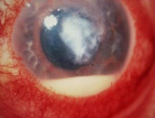
Corneal Ulcer is the deterioration of the external zone of the cornea. Corneal Ulcer is also known as Ulcerative keratitis or Eyesore. This is an inflammatory and infectious condition of the eye, which involves damage of one the layers of the cornea. This is usually seen in people living in tropical countries especially those involve in farming.

Corneal Ulcer in Dogs

The incidence of corneal ulcer among dogs normally happens when they pat or scratch their paws on their eye. Whenever it is scraped, the cornea becomes inflamed and become hazy. But this usually goes away without intervention.

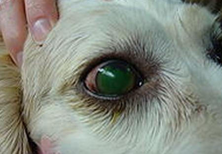
Corneal Ulcer in dogs

Picture 1 – Corneal Ulcer in Dogs

Corneal Ulcer Signs & Symptoms

This disorder usually present likes symptoms of infection. Signs and symptoms include:

  1. Bloodshot eyes
  2. Intense eye pain
  3. Lacrimation or tearing
  4. Clouded and frosty vision
  5. A disturbing sensation that seems like there is an object inside the eye
  6. Cannot look directly at bright lights due to intense pain
  7. Watery eyes
  8. Tingling sensation
  9. Dense drainage from the affected eye
  10. Inflamed eyelids
  11. Ball-shaped stain in the cornea, which is discernible even with the unaided eye

Corneal Ulcer Causes

The most frequent and usual cause of this disorder is infections by bacteria, parasites, virus or fungi. Others are caused by trauma, disorders of the eye, chemical contact, and some physical agents.

Bacterial causes. This is common among people who regularly use contact lenses either for aesthetic purposes or ophthalmologic purposes.

Viral causes. Virus is also one of the usual causes of this disorder. The most common virus causing this disorder is the herpes simplex virus. This virus can cause injury to the outer parts and on the most immersed part of the surface of the eye sometimes.

Fungal causes. Infections cause by fungi also results in corneal ulcer. This occurs due to inappropriate use of contact lenses and too much usage of steroidal eye drops. Fusarium is usually correlated with the outbreak of fungal keratitis seen among those who wear contact lens.

Parasitic causes. Parasites can also cause corneal ulcer. This is usually caused by the parasite acanthamoeba, which invade the eye through contact lens use. They are normally found in non-flowing forms of water like in the bath tubs, pools and water tap.

Other causes.

  1. Trauma to the eye can also result in corneal ulcer. This can either be due to accidental bumping of the eye, which results in scrapes and lacerations of the cornea. This insult allows the invasion of microorganisms into the eye causing infection.
  2. Eyes that have been dry for some time loses its natural barrier against trauma or infection.
  3. Chronic allergic reaction that affects the eye
  4. Accidental chemical spatter in the eye

Corneal Ulcer Diagnosis

Diagnosis for this corneal ulcer is usually made through direct scrutiny of the eye under an instrument to enlarge the contour of the eye. This procedure is known as slit lamp. Fluorescein staining allows better visualization of the cornea and will unveil condition of the cornea.

  1. Scraping a part of the cornea for examination under a microscope will reveal if bacteria or fungi is present.
  2. Culturing the corneal sample will also reveal specific microorganism.
  3. Blood work up is also done to see the presence of any inflammatory disorder.

Corneal Ulcer Treatment

Once the disease is properly diagnosed, specific treatment will be given. Your ophthalmologist will discuss management depending on how grave the disease and its cause.

Medications

1. Antibiotics. Antibiotic therapy is given to kill all microorganisms that can cause bacterial corneal ulcer. This is the immediate line of defense for corneal ulcer. Broad spectrum antimicrobials are mostly prescribed to contain all known pathogens.

  • Cefazolin: This antibiotic belongs to the first-generation class of cephalosporin. This drug specifically acts on gram-positive bacteria. It is commonly given in addition to aminoglycosides to attain extensive range of coverage. 50-133 mg/ml solution is usually added for better effect.
  • Gentamycin: This is an aminoglycoside antimicrobial agent that is specifically sensitive to gram-negative bacteria. Normally given in conjunction with cefazolin for better coverage.
  • Erythromycin: This antibiotic drug is prescribed for treating infections brought by predisposed group of bacteria. This is also given for interrupting infections to the cornea and conjunctiva.
  • Ophthalmic Ciprofloxacin: This drug is a bactericidal agent that prevents the production of bacterial DNA thereby stopping the development of the bacteria by preventing the DNA gyrase in predisposed individuals. This is also prescribed to patients infected with microorganisms that have become resistant to other antimicrobials.

2. Cycloplegics. These are agents that cause numbness of the ciliary muscles of the eye. They act to reduce the pain brought about by the twitching of eye muscles.

3. Ophthalmic Scopolamine: This drug works by obstructing the function of the agents that control muscle contraction, which makes the pupil of the eye enlarge, and cause numbness and paralysis of eye adjustment.

4. Antivirals. Antiviral therapy starts with mechanically removing the affected part of the cornea. After the removal, introduction of antiviral topical solutions.

  • Vidarabine: This drug acts to intervene with the first stages of the production of viruses. This is given if the patient develops an allergic reaction or cannot tolerate Idoxuridine.
  • Idoxuridine: This drug is initially prescribed for infections of the epithelium of the eye. It also deter the generation of herpes simples virus by making false DNA counterpart, which avert the virus from contaminating and impairing the tissues.

5. Antifungals. This line broad coverage antifungal produces the least sensation of pain and damage to cornea.

Natamycin: This antifungal drug is the drug of choice for curing infections cause by fungi in the cornea. This acts by binding the fungal cell membrane to form a convoluted and tangled compound, which changes the capacity of the membrane to allow passage and destroying important contents of the cells, killing the fungi. Therapy lasts for 14-21 days up until the desired result is achieved.

6. Adjunctive Drug Therapy. This therapy is needed to treat other conditions related to corneal ulcer or conditions brought about by the disease.

NSAIDs (Non-Steroidal Anti-Inflammatory Drugs). The action of NSAIDs in corneal ulcer is assumed to be related to the suppression of the enzyme prostaglandin. The production of this agent causes constriction and lessened vascular accessibility, increase in the white blood cells and pressure inside the eye.

  • Ibuprofen: This is regularly the drug chosen from this group. This is prescribed for the treatment of pain as long as it is not contraindicated to the patient. It acts by repress the reactions of the body to inflammation and pain by lowering the actions of the cyclooxegenase.
  • Analgesics. This agent is mainly given to provide comfort to the patient by reducing pain.
  • Acetaminophen: This is prescribed for mild to chronic pain.

Surgery

Corneal transplant only indicated when the disease cannot be treated with medications or if the medicines only worsen the condition.

Self Care

These are simple steps in caring for the eye to avoid or prevent the condition from getting worse.

  1. Remove contact lenses before going to bed.
  2. Wash your hands regularly especially before touching the eyes.
  3. Do not stroke the eye with your fingers.
  4. Place cold pack over the affected eye.

Corneal Ulcer Healing Time

There are two methods by which corneal ulcer heal, by movement of the epithelial cells around the area followed by division of the cells, or via addition of new blood vessel coming from the conjunctiva. Evident tiny ulcers improve very fast by the first approach. Those ulcers, which are bigger and more profound, recover via the second approach. The white blood cells and fibroblasts helps heal the ulcers. They provide new tissues that completely heal the cornea. Normally, corneal ulcers resolve by the end of the fourth day.

Corneal Ulcer Pictures

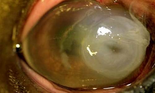
Here are the some pictures on corneal ulcer

Corneal Ulcer pictures 3

Picture 2 – Starting stage of corneal Ulcer (easily treated)

Corneal Ulcer pictures 5

Corneal Ulcer pictures 1

Corneal Ulcer pictures

Picture 5 – Very Severe stage (observe thinning of cornea). Need Corneal Transplant

No responses yet

Blocked Tear Duct in Babies and Adults – Treatment, Symptoms, Pictures

Mar 09 2012 Published by under Diseases & Conditions

What is Blocked Tear Duct?

When the small tubes or ducts that drain tears from the eyes are obstructed, the resulting condition is a blocked tear duct; this is also called as dacryostenosis.

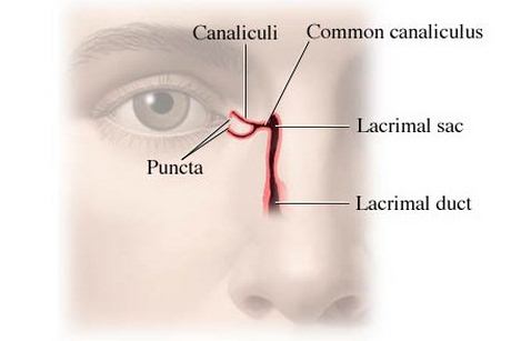
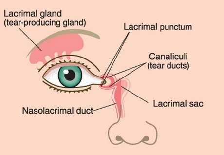
The Anatomy of Tear Ducts

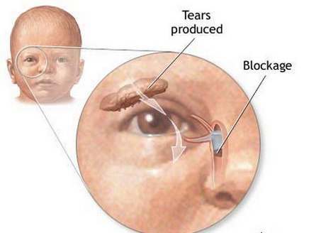
The tear duct or the nasolacrimal duct is a structure responsible for draining tears from the lacrimal gland or sac to the nasal cavity. This duct runs through the eyes and into the nasal cavity, it actually drains some of the excess tears into the inferior nasal meatus. This is the reason why crying is followed or accompanied by a runny nose.

The tear ducts have a small opening in the inner canthus of the eye (inner part of the eye), acquired obstructions can plug this opening and the drainage of the nasolacrimal ducts. Aside from that, congenital conditions, tumors and infections also contribute to a blocked tear duct.

Anatomy of lacrimal apparatus

Image Source – Health.com

canaliculi, Nasolacrimal duct & Lacrimal apparatus

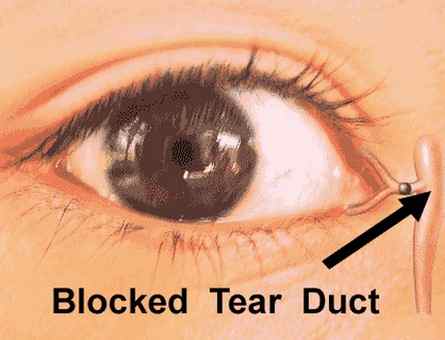
Picture 1,2  shows lacrimal duct, lacrimal glands and tear ducts

Blocked Tear Duct in Adults

Dacryostenosis is a common occurrence among newborns or pediatric clients. But the same condition can also affect adults. According to certain studies, almost 3% of clinical visits are due to problems of the nasolacrimal duct. In adults, probable causes of an obstruction of the tear ducts include the following:

  1. Chronic inflammation
  2. Fibrosis
  3. Neoplasms or malignant growths
  4. Infections
  5. Trauma
  6. Systemic diseases

Chronic Inflammation

Inflammatory processes can cause the tear ducts to be blocked. Chronic inflammation is more commonly seen in adults than in children. Such inflammation can be caused by other underlying diseases such as an autoimmune disease.

Fibrosis

Fibrosis formation is usually a result of chronic inflammation or infection. Fibrosis is the formation of fibrotic tissues, which are hard and alters the structure of the adjacent healthy tissues. Fibrous formations can be compared to scars. When scars form in the nasolacrimal duct (which is relatively small area), obstructions can occur.

Neoplasms or Malignant Growths

Malignant growths that obstruct the nasolacrimal gland are rare occurrences but is life threatening. Neoplasms or malignant tumors usually arise in the epithelial or mesenchymal layer of the lacrimal gland, where epithelial tumors account for almost 70% of the cases. Most malignancies affecting the nasolacrimal duct is a manifestation of another systemic condition.

Infections

Bacterial or fungal infections in the eye and nasolacrimal duct can also cause obstructions. Debris from the microorganism and the body cells that fight these organisms can block the small tubes and openings of the nasolacrimal drainage. Microorganisms such as Staphyloccoccus aureaus, Streptococcus, Chlamydia and even Helicobacter pylori are related to infectious obstructions of the tear duct.

Systemic Diseases

Systemic conditions such as sarcoidosis and Wegners granulomatosis can also be causes of bloked tear ducts. Systemic diseases are conditions that affect the whole body and not just a local organ or system. In both conditions, inflammatory cells or granulomas form in different parts of the body for unknown reasons.

blokage of tear ducts

Blocked Tear Duct pictures

Picture 3, 4 shows blockage of tear duct system

Image Source – Medindia.net

Blocked Tear Ducts in Babies

Blocked tear ducts are most common among newborns and infants than in adults and it has the following causes:

  1. Congenital malformations
  2. Malformations of the skull and face
  3. Facial injuries
  4. Infections or inflammations

Congenital malformations

A blocked tear duct occurs in almost 6 out of 100 babies, or almost 20% among newborns in some studies. This occurs primarily due to the nasolacrimal duct that formed abnormally (begins inside the womb), or a tear duct that is not yet fully developed.

In most cases a flap of thin tissue covers the opening of the tear duct, as the new born grows and develops, the flap of tissue disappears and the opening becomes patent.

However, in certain conditions where the tear ducts fail to develop and remain blocked, medical treatment and surgery might be considered.

Malformations of the skull and face

An abnormally developed nasal bone can press onto the tear duct, closing if off and leading to an obstruction. Conditions where the nasal cavity and other structures of the face are malformed can also cause a blocked tear duct.

Infections or inflammations

Cellular debris from bacteria and white blood cells, as well as the swelling brought about by the inflammatory process can cramp up the nasolacrimal duct causing it to be obstructed.

Symptoms

  1. Wet teary eyes
  2. Excessive tearing or epiphora
  3. Redness of the eye
  4. Puffy eyes
  5. Pain or swelling of the lacrimal glands (found at the inner corner of the eye)
  6. Pus discharge from the eye or lacrimal gland
  7. Eyelid inflammation
  8. Vision changes
  9. Presence of cyst or tumor on the corner of the eye
  10. Asymmetrical eyes
  11. Unilateral exophthalmia or bulging of the eyes
  12. Presence of multiple granulomas surrounding the eye
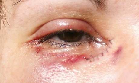
Blocked Tear Ducts external appearance image
Picture 5 – External appearance in Blocked Tear Ducts condition

Causes

  1. Congenital malformations
  2. Craniofacial abnormalities
  3. Chronic inflammation
  4. Infection
  5. Tumor growths or neoplasms
  6. Facial injuries or trauma
  7. Systemic conditions

Diagnosis

To diagnose a blocked tear duct, the following tests are done:

Dacryocystoshraphy

This is an eye imaging test where a contrast dye is utilized to evaluate the drainage of the nasolacrimal duct. The contrast dye is passed through the patient’s tear drainage system and a CT scan, MRI or an X-Ray is taken while the dye is inside the nasolacrimal duct to determine the cause of obstruction and its location.

Fluoresceine dye disappearance test

In this test a drop of a special dye is placed on the eyes. With a normal tear duct, some of the dye would be absorbed and drained by our tear ducts, but when a significant amount stays on your eye, it could indicate an obstruction.

Irrigation

This procedure simply involves irrigation of the eye with a saline solution to check if the ducts are draining properly.

Probing

A small, slender probe is inserted in the puncta (the small opening on the corner of the eye), to view the nasolacrimal duct and check for any obstructions.

Treatment

Treatment can be classified as Invasive and minimally invasive procedures

Minimally invasive procedures

Topical Antibiotics and Massage

This treatment regimen is particularly effective to younger age groups (1-12 months old) with blocked tear ducts. Proper massage can stimulate the drainage of the tear ducts and improve the child’s condition. In a study, almost 32% of the patients with blocked tear ducts improved with the two interventions alone.

Irrigation and Probing

The two procedures are usually done to diagnose the condition or figure out the cause of the obstruction. In certain cases (especially for pediatric patients), irrigation and probing can serve as the treatment for the condition. This may be due to the fact that the procedure dilates the puncta of the eye, which the relives the condition in most cases.

Use of Crawford Tubes

Insertion of tubes to dilate the nasolacrimal duct had been proven effective for children of all age groups. This treatment is a surgical treatment and is done if massage, antibiotics and probing failed to relieve the condition.

Invasive Treatment

External Dacryocystorhinostomy

This is a surgical procedure that involves the creation of a fistula of the lacrimal gland into the nasal cavity. This can alleviate the symptoms such as poor drainage and inflammation. This procedure was first done in 1911, but has failed due to unwanted side effects. But with the advances in medical and surgical technology, the procedure approaches a nearly 100% success rate.

Complications

When an obstructed tear duct is not treated, it could lead to the following complications:

  1. Recurrent infections
  2. Pain and discomfort brought about by the swelling
  3. Stagnant drainage
  4. Chronic inflammation
  5. Conjunctivitis
  6. Blepharitis
  7. Severe exophthalmia (in the case of tumor fomations)

Seek immediate Medical Attention if:

  1. There is a cyst, tumor or growth that is large or rapidly growing
  2. If the obstruction causes severe and persistent symptoms
  3. If the obstruction causes changes in the contour, shape and position of the eyes
  4. If other systemic symptoms are present, such as fever, pain, fatigue, and weakness

Prevention

Prevention primarily focuses on measures to avoid eye infection or inflammation. Other causes of blocked tear ducts such as congenital anomalies have no known prevnetions.

Prevent a blocked tear duct by employing the following measures:

  1. Avoid contact with people who have eye infections such as conjunctivitis
  2. Do not rub your eyes especially with bare hands or if your hands are not clean
  3. Proper hygiene and make up removal
  4. Keep contact lenses clean, use them properly and hygienically
  5. Wash your hand regularly

No responses yet

Swollen Uvula – Symptoms, Causes, Treatment and Remedies

Mar 06 2012 Published by under Diseases & Conditions

What is Swollen Uvula?

Swollen Uvula is also termed by the medical professionals as Uvilitis. It just means that your uvula is inflamed. Uvula is a tissue that is hanging back in our mouth that aids in food passageway for it will not go to our nasal cavity that aids in our breathing and it helps in the sound we create when we are speaking. Having this kind of disease makes a person irritable and uncomfortable especially when eating, drinking and even just swallowing the saliva.

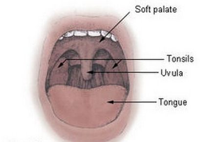
Uvula Location & surroundings

Picture 1 – Uvula Location & its surroundings structures

Signs and Symptoms

  1. Dry mouth and throat due to the inflammation experiencing by the person who has a swollen uvula even though that person drinks water there will be a feeling of dryness.
  2. Sore Throat due to infection or irritation that can contribute in having swollen uvula.
  3. Headache due to the irritation in the uvula.
  4. Throat Irritation can irritate the uvula and it can lead in inflammation
  5. There is a feeling to vomit because the irritation of the uvula the gag reflex is stimulated.
  6. Difficulty in Swallowing because of the irritation cause by the uvula.
  7. Roughness of voice because the uvula and throat is affected that aids us when we speak.
  8. Fever because of the infection that is present in the swollen uvula.
  9. Pus discharge is due to prolong unaided infection.
  10. Desire to cough due to the irritation that the person feels.
Swollen Uvula (enlarged uvula) Pictures
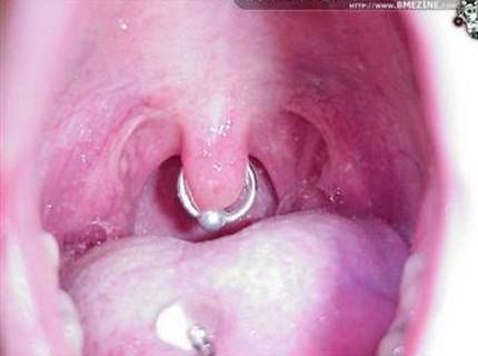
Picture 2 – Observe the red, enlarged Uvula in Swollen Uvula condition

Causes

There are different reasons that might lead in uvula swelling. These are the common causes for having swollen uvula:

  1. Bacterial or Viral Infection can lead to inflammation and soreness of the throat and the surrounding tissues.
  2. Dehydration is one of the common causes for having a swollen uvula. It is where the body only gets a little amount of liquid and the uvula is soak in saliva.
  3. Excessive smoking and inhalation of different pollutants that can lead to dryness of the uvula as well as it makes the uvula swell and became itchy.
  4. Allergies that will cause the uvula to swell.
  5. Throat diseases like common cold and sore throat can make the uvula inflamed.
  6. Trauma that can cause swelling of the uvula like eating spicy and hot substance, drinking of alcohol, and instruments that are inserted through mouth like endoscope.
  7. When the person is snoring because it leads to the malfunctioning or changes in breathing pattern which also affects the uvula is one of the reasons why the uvula is swollen.
  8. Drinking too much alcohol can cause dehydration as well as it leads to snoring that can cause swelling and irritation.

Diagnosis

Medical experts diagnose if the person has a swollen uvula according to presenting signs and symptoms, by doing physical assessment to the patient.

(1) Medical History of the patient and

(2) Culture and Sensitivity to check for presence of infection in the uvula.

Remedies

There are different ways that can be done in treating the swollen uvula. This disease is not a life-threatening and a serious problem this condition can be cured within 24 hours. These remedies are:

  1. Drinking water is one of the best remedies to treat swollen uvula. Dehydration is one of the causes of having the disease, so, to cure dehydration drinking water is one of the effective remedies to treat swollen uvula.
  2. Honey or Aloe Vera Juice is also known for treating swollen uvula.
  3. Eating garlic cloves and onion can help to cure the swollen uvula because of its anti-bacterial property and anti-inflammatory properties.
  4. Adding salt in the drinking water can help in curing throat irritation and inflammation.
  5. Cold drinks and beverages can also help in alleviating the irritation feeling caused by swollen uvula. You can use this kind of remedy if you do not have a cold.
  6. Gargle using the mixture of salt and water can help to treat swollen uvula by reducing the pain, irritation and swelling.
  7. Drinking tea with basil leaves is also known for the treatment of swelling of the uvula.
  8. Mix turmeric with cold or warm water. Turmeric is known for anti-inflammatory healing properties, so, it’s a good cure for swollen uvula that can relieve swelling and irritation.

Treatment

Swollen Uvula normally doesn’t need a special treatment because it can cure at home but there are some instances that home remedies don’t help to cure or alleviate the Swollen Uvula. Go to the Otalaryngologist which are experts in this disease. If the swollen uvula doesn’t cure within 24-48 hours it is the best time to cure the swollen uvula by the medical professionals. This disease is not a life-threatening type but if you are diagnosed with this type of disease it is advised to seek immediate attention to avoid formation of other disease due to having Swollen Uvula.

  1. Medication to alleviate pain and swelling like Aspirin that can help to ease pain and swelling as well as to reduce fever.
  2. When there is a presence of bacterial or viral infection the doctor can give antibiotics to cure infection.
  3. Doctors also advised the patients to change some lifestyle habits like drinking alcohol as well as smoking because they can lead to irritation as well as infection and it can weaken the immune system of the body.
  4. If this disease recurs time to time normally the doctors advised the patients to have Adrenalin shots. In this disease, where the uvula is swollen the air passages constricts when given this shots it will help to reduce swelling as well as to reduce irritation caused by this disease that recurs every time.

Complications

There are no related specific complication having a swollen uvula because it’s not a severe problem that can harm any person having this kind of disease and if it’s treated as early as it’s diagnosed there will be no specific problem. These complications arise only if the person doesn’t seek immediate medical attention.

(1) Necrosis of the uvula that will lead of having a surgery that will remove the uvula. Removing the uvula can be a problem because of its function in the human system, as mentioned earlier uvula helps as when we eat foods and it helps us when we speak, so, removing it means the person will have difficulty when eating as well as when speaking.

(2) Severe infection because of the delayed seeking medical treatment. To avoid these two complications, it is advised that the person must seek medical attention if home remedies don’t became effective in treating swollen uvula.

No responses yet

High Grade Squamous Intraepithelial Lesion – Symptoms, Causes, Treatment

Mar 02 2012 Published by under Diseases & Conditions

What is High Grade Squamous Intraepithelial Lesion (HGSIL)?

High grade squamous intraepithelial lesion or HSIL is a finding on the cervical tissues following a Pap smear. HSIL is a type of cervical dysplasia found in microscopic analysis of the cervical cells. Cervical dysplasia refers to the occurrence of pre-malignant or precancerous cells in the cervix and opening of the uterus. With this regard, HSIL indicates a moderate dysplasia to severe neoplasia of the cervical cells that may mean carcinoma in situ.

HSIL is different from low grade squamous intraepithelial lesion or LSIL in which the former have more defined changes in the shape and size of the cervical cells. LSIL only involve the appearance of early changes in the shape and size and is often associated with the presence of human papilloma virus or genital warts.

High grade squamous intraepithelial lesion may eventually lead to invasive cancer of the cervix when managements are not instituted. Being carcinoma in situ, it is considered stage 0 cervical cancer, but may not indicate that there is already cervical cancer. The presence of HSIL should be treated to prevent the development into cervical cancer.
High grade squamous intraepithelial neoplasia can be seen in various areas such as the esophagus, cervix, vagina and vulva, where there are squamous epitheliums.

Any detection of HSIL requires further tests to evaluate presence of cancer. Only 2% of patients with HSIL have invasive cancer, however up to 20% with HSIL will develop cancer if it is left untreated. Prevention of HSIL developing into cancer involves removing or destroying the precancerous cells.

Symptoms of HSIL

The appearance of cervical dysplasia such as the high grade squamous intraepithelial lesion usually does not result in signs and symptoms. The only manifestation of the condition is the microscopic changes that happen in the cervical cells evident upon microscopic examination. When HSIL occurs along with cervical cancer, there are symptoms associated with the cervical malignancy such as

  1. pain on the area
  2. bleeding after intercourse
  3. vaginal bleeding and
  4. abnormal vaginal secretions

Causes of HSIL

The cause of any type of cervical dysplasia such as the high grade squamous intraepithelial lesion is similar to the risk factors for cervical cancer, which includes:

Human Papilloma Virus

Infection with HPV or genital warts can lead to HSIL. This is spread through sexual contact and is one of the most common sexually transmitted diseases because of being asymptomatic sometimes. HPV usually affects young women aged 15 to 25 years old. Most women become infected with HPV without them knowing. HPV usually resolves spontaneously without any treatment, but it may have cause cervical dysplasia by that time if not treated. Recurrent HPV infection is commonly associated with cervical dysplasia and cervical cancer. Since HPV is transmitted through sexual contact, preventing it involves having only one sex partner and ensuring safe sex practices all the time. Increased risk for HPV infection involves having a reduced immune system or smoking.

Diagnosis of HSIL

Diagnostic Tests for high grade squamous intraepithelial lesion involves undergoing Pap smear. Pap smear or Pap test is done during pelvic examination where a speculum is inserted to collect samples of the cervical mucus. The mucus is then smeared and examined under the microscope. High grade squamous intraepithelial dysplasia of the cervix is then observed as the type of cervical dysplasia happening on the cervical cells.

When HSIL is observed, further tests are done to evaluate the presence of cervical cancer. These include:

Colposcopy

Colposcopy involves the use of a microscope or colposcope to study or visualize the cervix. Colposcopy helps identify specific areas of cervical dysplasia and can check the totality of the cervix. The abnormal cells may be removed at the time of colposcopy to prevent further spread. This management is called “see and treat”. Colposcopy may last for one hour and results are made depending on the extent of abnormality.

Biopsy

When suspicious areas are seen during colposcopy, a biopsy may be done to check if the cells are benign or malignant. Abnormal dysplasia in the cervix as seen in biopsies is termed cervical intraepithelial neoplasia (CIN). CIN is further classified into:

  1. CIN 1– This involves the presence of dysplasia on 1/3 portion of the cervical epithelium
  2. CIN 2– This involves the presence of dysplasia on the 2/3 portion of the cervical lining and is a more serious cervical dysplasia
  3. CIN 3– This can be categorized as carcinoma in situ in which the dysplasia affects more than 2/3 of the cervical lining.
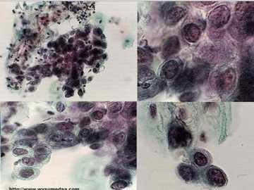
biopsy results in High Grade Squamous Intraepithelial Lesion
Biopsy results in High Grade Squamous Intraepithelial Lesion

HPV testing

HPV testing is also performed in women to detect presence of HPV infection.

Treatment of HSIL

The presence of cervical intraepithelial neoplasia in biopsy may prompt for treatments. Treatments include:

Loop Electrosurgical Excision Procedure or LEEP

This surgical procedure involves the excision of abnormal cells through the introduction of electric current through a wire loop in the cervix. When high grade squamous intraepithelial lesion is diagnosed in pregnant women, LEEP may be postponed and done after delivery because it can cause premature labor or spontaneous abortion in the first trimester. Progression of HSIL is usually slow during pregnancy so LEEP may be done later.

Conization

Conization is another approach in the treatment of HSIL. This involves the removal of a cone-shaped tissue from the cervix to get the deeper layers of abnormal cells and not just the superficial cervical lining. This is usually done during biopsy and is also called cone biopsy.

Cryotherapy

This surgical procedure involves the use of very cold substances to freeze the abnormal cervical cells. After freezing, the area exposed is usually removed.

Laser Therapy

This procedure involves the use of laser beams to destroy and remove the abnormal cells.

Follow Up Care

After the removal or the excision of the abnormal cervical cells, follow-up check-ups are needed to ascertain that no cervical cancer or another HSIL is taking place. Cells may still become abnormal, despite the surgical removal and may progress to cervical cancer when not detected early. Follow-up care includes:

  • Pap smear with colposcopy every 6 months in one year followed by annual pap smears when there are no abnormal cells seen.
  • Pap smear and colposcopy every 6 months when abnormal cells are seen after the first year of follow-up.

Prognosis of HSIL

The presence of HSIL should not be mistaken as presence of cancer. HSIL has a good prognosis when treatments are instituted. In fact, only 20% of cases progresses to cervical cancer without treatment, which means that early management for HSIL will not eventually lead to cervical malignancy.

Finally, Compare with – Low Grade Squamous Intraepithelial Lesion – Symptoms, Causes, Treatment 

No responses yet

« Prev

© 2017 eHealthWall.com. All Rights Reserved. Privacy Policy
This website is for informational purposes only and Is not a substitute for medical advice, diagnosis or treatment.